4MBX

Structure of unliganded B-Lymphotropic Polyomavirus VP1


Experimental Data Snapshot

  • Method: X-RAY DIFFRACTION
  • Resolution: 1.92 Å
  • R-Value Free: 0.198 
  • R-Value Work: 0.165 
  • R-Value Observed: 0.167 

wwPDB Validation   3D Report Full Report


This is version 1.2 of the entry. See complete history


Literature

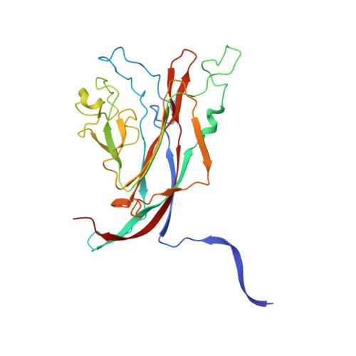
Structures of B-Lymphotropic Polyomavirus VP1 in Complex with Oligosaccharide Ligands.

Neu, U.Khan, Z.M.Schuch, B.Palma, A.S.Liu, Y.Pawlita, M.Feizi, T.Stehle, T.

(2013) PLoS Pathog 9: e1003714-e1003714

  • DOI: https://doi.org/10.1371/journal.ppat.1003714
  • Primary Citation of Related Structures:  
    4MBX, 4MBY, 4MBZ

  • PubMed Abstract: 

    B-Lymphotropic Polyomavirus (LPyV) serves as a paradigm of virus receptor binding and tropism, and is the closest relative of the recently discovered Human Polyomavirus 9 (HPyV9). LPyV infection depends on sialic acid on host cells, but the molecular interactions underlying LPyV-receptor binding were unknown. We find by glycan array screening that LPyV specifically recognizes a linear carbohydrate motif that contains α2,3-linked sialic acid. High-resolution crystal structures of the LPyV capsid protein VP1 alone and in complex with the trisaccharide ligands 3'-sialyllactose and 3'-sialyl-N-acetyl-lactosamine (3SL and 3SLN, respectively) show essentially identical interactions. Most contacts are contributed by the sialic acid moiety, which is almost entirely buried in a narrow, preformed cleft at the outer surface of the capsid. The recessed nature of the binding site on VP1 and the nature of the observed glycan interactions differ from those of related polyomaviruses and most other sialic acid-binding viruses, which bind sialic acid in shallow, more exposed grooves. Despite their different modes for recognition, the sialic acid binding sites of LPyV and SV40 are half-conserved, hinting at an evolutionary strategy for diversification of binding sites. Our analysis provides a structural basis for the observed specificity of LPyV for linear glycan motifs terminating in α2,3-linked sialic acid, and links the different tropisms of known LPyV strains to the receptor binding site. It also serves as a useful template for understanding the ligand-binding properties and serological crossreactivity of HPyV9.


  • Organizational Affiliation

    Interfaculty Institute of Biochemistry, University of Tuebingen, Tuebingen, Germany.


Macromolecules
Find similar proteins by:  (by identity cutoff)  |  3D Structure
Entity ID: 1
MoleculeChains Sequence LengthOrganismDetailsImage
Major Capsid Protein VP1
A, B, C, D, E
A, B, C, D, E, F, G, H, I, J
278B-lymphotropic polyomavirusMutation(s): 0 
Gene Names: VP1
UniProt
Find proteins for P04010 (B-lymphotropic polyomavirus)
Explore P04010 
Go to UniProtKB:  P04010
Entity Groups  
Sequence Clusters30% Identity50% Identity70% Identity90% Identity95% Identity100% Identity
UniProt GroupP04010
Sequence Annotations
Expand
  • Reference Sequence
Small Molecules
Ligands 2 Unique
IDChains Name / Formula / InChI Key2D Diagram3D Interactions
EDO
Query on EDO

Download Ideal Coordinates CCD File 
AA [auth E]
BA [auth E]
DA [auth F]
EA [auth F]
FA [auth F]
AA [auth E],
BA [auth E],
DA [auth F],
EA [auth F],
FA [auth F],
GA [auth F],
IA [auth G],
JA [auth G],
KA [auth G],
L [auth A],
LA [auth G],
M [auth A],
MA [auth G],
N [auth A],
OA [auth H],
P [auth B],
Q [auth B],
QA [auth I],
RA [auth I],
S [auth C],
TA [auth J],
U [auth D],
UA [auth J],
V [auth D],
X [auth E],
Y [auth E],
Z [auth E]
1,2-ETHANEDIOL
C2 H6 O2
LYCAIKOWRPUZTN-UHFFFAOYSA-N
CA
Query on CA

Download Ideal Coordinates CCD File 
CA [auth F]
HA [auth G]
K [auth A]
NA [auth H]
O [auth B]
CA [auth F],
HA [auth G],
K [auth A],
NA [auth H],
O [auth B],
PA [auth I],
R [auth C],
SA [auth J],
T [auth D],
W [auth E]
CALCIUM ION
Ca
BHPQYMZQTOCNFJ-UHFFFAOYSA-N
Experimental Data & Validation

Experimental Data

  • Method: X-RAY DIFFRACTION
  • Resolution: 1.92 Å
  • R-Value Free: 0.198 
  • R-Value Work: 0.165 
  • R-Value Observed: 0.167 
  • Space Group: C 1 2 1
Unit Cell:
Length ( Å )Angle ( ˚ )
a = 150.01α = 90
b = 95.92β = 95.82
c = 231.68γ = 90
Software Package:
Software NamePurpose
MxCuBEdata collection
MOLREPphasing
REFMACrefinement
XDSdata reduction
XSCALEdata scaling

Structure Validation

View Full Validation Report


Entry History 

Deposition Data

Revision History  (Full details and data files)

  • Version 1.0: 2013-12-04
    Type: Initial release
  • Version 1.1: 2014-01-22
    Changes: Source and taxonomy
  • Version 1.2: 2023-09-20
    Changes: Data collection, Database references, Derived calculations, Refinement description