Towards accurate structural characterization of metal centres in protein crystals: the structures of Ni and Cu T6 bovine insulin derivatives.
Frankaer, C.G., Mossin, S., Stahl, K., Harris, P.(2014) Acta Crystallogr D Biol Crystallogr 70: 110-122
- PubMed: 24419384
- DOI: https://doi.org/10.1107/S1399004713029040
- Primary Citation of Related Structures:
4M4F, 4M4H, 4M4I, 4M4J, 4M4L, 4M4M - PubMed Abstract:
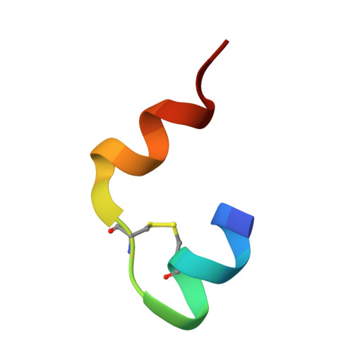
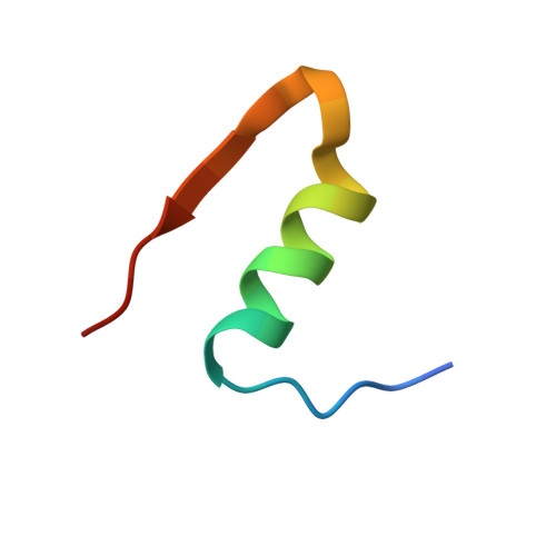
Using synchrotron radiation (SR), the crystal structures of T6 bovine insulin complexed with Ni(2+) and Cu(2+) were solved to 1.50 and 1.45 Å resolution, respectively. The level of detail around the metal centres in these structures was highly limited, and the coordination of water in Cu site II of the copper insulin derivative was deteriorated as a consequence of radiation damage. To provide more detail, X-ray absorption spectroscopy (XAS) was used to improve the information level about metal coordination in each derivative. The nickel derivative contains hexacoordinated Ni(2+) with trigonal symmetry, whereas the copper derivative contains tetragonally distorted hexacoordinated Cu(2+) as a result of the Jahn-Teller effect, with a significantly longer coordination distance for one of the three water molecules in the coordination sphere. That the copper centre is of type II was further confirmed by electron paramagnetic resonance (EPR). The coordination distances were refined from EXAFS with standard deviations within 0.01 Å. The insulin derivative containing Cu(2+) is sensitive towards photoreduction when exposed to SR. During the reduction of Cu(2+) to Cu(+), the coordination geometry of copper changes towards lower coordination numbers. Primary damage, i.e. photoreduction, was followed directly by XANES as a function of radiation dose, while secondary damage in the form of structural changes around the Cu atoms after exposure to different radiation doses was studied by crystallography using a laboratory diffractometer. Protection against photoreduction and subsequent radiation damage was carried out by solid embedment of Cu insulin in a saccharose matrix. At 100 K the photoreduction was suppressed by ∼15%, and it was suppressed by a further ∼30% on cooling the samples to 20 K.
Organizational Affiliation:
Department of Chemistry, Technical University of Denmark, Kemitorvet 207, DK-2800 Kgs. Lyngby, Denmark.