4M1B

Structural Determination of BA0150, a Polysaccharide Deacetylase from Bacillus anthracis


Experimental Data Snapshot

  • Method: X-RAY DIFFRACTION
  • Resolution: 1.99 Å
  • R-Value Free: 0.214 
  • R-Value Work: 0.170 
  • R-Value Observed: 0.172 

wwPDB Validation   3D Report Full Report


This is version 1.2 of the entry. See complete history


Literature

Structure determination of BA0150, a putative polysaccharide deacetylase from Bacillus anthracis.

Strunk, R.J.Piemonte, K.M.Petersen, N.M.Koutsioulis, D.Bouriotis, V.Perry, K.Cole, K.E.

(2014) Acta Crystallogr Sect F Struct Biol Cryst Commun 70: 156-159

  • DOI: https://doi.org/10.1107/S2053230X13034262
  • Primary Citation of Related Structures:  
    4M1B

  • PubMed Abstract: 

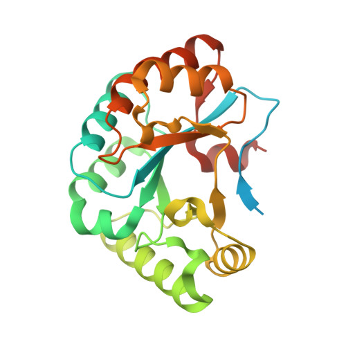
    Polysaccharide deacetylases are bacterial enzymes that catalyze the deacetylation of acetylated sugars on the membranes of Gram-positive bacteria, allowing them to be unrecognized by host immune systems. Inhibition of these enzymes would disrupt such pathogenic defensive mechanisms and therefore offers a promising route for the development of novel antibiotic therapeutics. Here, the first X-ray crystal structure of BA0150, a putative polysaccharide deacetylase from Bacillus anthracis, is reported to 2.0 Å resolution. The overall structure maintains the conserved (α/β)8 fold that is characteristic of this family of enzymes. The lack of a catalytic metal ion and a distinctive metal-binding site, however, suggest that this enzyme is not a functional polysaccharide deacetylase.


  • Organizational Affiliation

    Department of Chemistry, Ithaca College, 953 Danby Road, Ithaca, NY 14850, USA.


Macromolecules
Find similar proteins by:  (by identity cutoff)  |  3D Structure
Entity ID: 1
MoleculeChains Sequence LengthOrganismDetailsImage
Polysaccharide deacetylase
A, B
254Bacillus anthracisMutation(s): 0 
Gene Names: ba0150BAS0150BA_0150GBAA_0150
UniProt
Find proteins for A0A6L7HRJ6 (Bacillus anthracis)
Explore A0A6L7HRJ6 
Go to UniProtKB:  A0A6L7HRJ6
Entity Groups  
Sequence Clusters30% Identity50% Identity70% Identity90% Identity95% Identity100% Identity
UniProt GroupA0A6L7HRJ6
Sequence Annotations
Expand
  • Reference Sequence
Small Molecules
Ligands 1 Unique
IDChains Name / Formula / InChI Key2D Diagram3D Interactions
PGE
Query on PGE

Download Ideal Coordinates CCD File 
C [auth B]TRIETHYLENE GLYCOL
C6 H14 O4
ZIBGPFATKBEMQZ-UHFFFAOYSA-N
Experimental Data & Validation

Experimental Data

  • Method: X-RAY DIFFRACTION
  • Resolution: 1.99 Å
  • R-Value Free: 0.214 
  • R-Value Work: 0.170 
  • R-Value Observed: 0.172 
  • Space Group: P 21 21 21
Unit Cell:
Length ( Å )Angle ( ˚ )
a = 47.173α = 90
b = 75.795β = 90
c = 124.54γ = 90
Software Package:
Software NamePurpose
ADSCdata collection
Rosettamodel building
PHENIXrefinement
XDSdata reduction
XDSdata scaling
Rosettaphasing

Structure Validation

View Full Validation Report


Entry History 

Deposition Data

Revision History  (Full details and data files)

  • Version 1.0: 2014-02-12
    Type: Initial release
  • Version 1.1: 2014-09-24
    Changes: Database references
  • Version 1.2: 2024-02-28
    Changes: Data collection, Database references, Derived calculations