4LXC

The antimicrobial peptidase lysostaphin from Staphylococcus simulans


Experimental Data Snapshot

  • Method: X-RAY DIFFRACTION
  • Resolution: 3.50 Å
  • R-Value Free: 0.287 
  • R-Value Work: 0.268 
  • R-Value Observed: 0.269 

wwPDB Validation   3D Report Full Report


This is version 1.3 of the entry. See complete history


Literature

Crystal structure of the antimicrobial peptidase lysostaphin from Staphylococcus simulans.

Sabala, I.Jagielska, E.Bardelang, P.T.Czapinska, H.Dahms, S.O.Sharpe, J.A.James, R.Than, M.E.Thomas, N.R.Bochtler, M.

(2014) FEBS J 281: 4112-4122

  • DOI: https://doi.org/10.1111/febs.12929
  • Primary Citation of Related Structures:  
    4LXC, 4QP5, 4QPB

  • PubMed Abstract: 

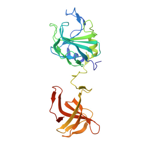
    Staphylococcus simulans biovar staphylolyticus lysostaphin efficiently cleaves Staphylococcus aureus cell walls. The protein is in late clinical trials as a topical anti-staphylococcal agent, and can be used to prevent staphylococcal growth on artificial surfaces. Moreover, the gene has been both stably engineered into and virally delivered to mice or livestock to obtain resistance against staphylococci. Here, we report the first crystal structure of mature lysostaphin and two structures of its isolated catalytic domain at 3.5, 1.78 and 1.26 Å resolution, respectively. The structure of the mature active enzyme confirms its expected organization into catalytic and cell-wall-targeting domains. It also indicates that the domains are mobile with respect to each other because of the presence of a highly flexible peptide linker. The high-resolution structures of the catalytic domain provide details of Zn(2+) coordination and may serve as a starting point for the engineering of lysostaphin variants with improved biotechnological characteristics.


  • Organizational Affiliation

    International Institute of Molecular and Cell Biology, Warsaw, Poland.


Macromolecules
Find similar proteins by:  (by identity cutoff)  |  3D Structure
Entity ID: 1
MoleculeChains Sequence LengthOrganismDetailsImage
Lysostaphin
A, B, C, D
255Staphylococcus simulansMutation(s): 0 
Gene Names: lssU66883.1
EC: 3.4.24.75
UniProt
Find proteins for P10547 (Staphylococcus simulans)
Explore P10547 
Go to UniProtKB:  P10547
Entity Groups  
Sequence Clusters30% Identity50% Identity70% Identity90% Identity95% Identity100% Identity
UniProt GroupP10547
Sequence Annotations
Expand
  • Reference Sequence
Small Molecules
Ligands 2 Unique
IDChains Name / Formula / InChI Key2D Diagram3D Interactions
SO4
Query on SO4

Download Ideal Coordinates CCD File 
F [auth A]
G [auth A]
I [auth B]
J [auth B]
K [auth B]
F [auth A],
G [auth A],
I [auth B],
J [auth B],
K [auth B],
N [auth D],
O [auth D],
P [auth D],
Q [auth D]
SULFATE ION
O4 S
QAOWNCQODCNURD-UHFFFAOYSA-L
ZN
Query on ZN

Download Ideal Coordinates CCD File 
E [auth A],
H [auth B],
L [auth C],
M [auth D]
ZINC ION
Zn
PTFCDOFLOPIGGS-UHFFFAOYSA-N
Experimental Data & Validation

Experimental Data

  • Method: X-RAY DIFFRACTION
  • Resolution: 3.50 Å
  • R-Value Free: 0.287 
  • R-Value Work: 0.268 
  • R-Value Observed: 0.269 
  • Space Group: P 43 3 2
Unit Cell:
Length ( Å )Angle ( ˚ )
a = 282.013α = 90
b = 282.013β = 90
c = 282.013γ = 90
Software Package:
Software NamePurpose
CBFlibdata collection
PHASERphasing
REFMACrefinement
XDSdata reduction
XSCALEdata scaling

Structure Validation

View Full Validation Report


Entry History 

Deposition Data

Revision History  (Full details and data files)

  • Version 1.0: 2014-07-09
    Type: Initial release
  • Version 1.1: 2014-07-16
    Changes: Other
  • Version 1.2: 2014-10-01
    Changes: Database references
  • Version 1.3: 2023-09-20
    Changes: Data collection, Database references, Derived calculations, Refinement description