4L9P

Crystal structure of Aspergillus fumigatus protein farnesyltransferase complexed with the FII analog, FPT-II, and the KCVVM peptide


Experimental Data Snapshot

  • Method: X-RAY DIFFRACTION
  • Resolution: 1.45 Å
  • R-Value Free: 0.152 
  • R-Value Work: 0.125 
  • R-Value Observed: 0.126 

wwPDB Validation   3D Report Full Report


Ligand Structure Quality Assessment 


This is version 1.3 of the entry. See complete history


Literature

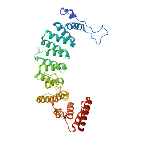
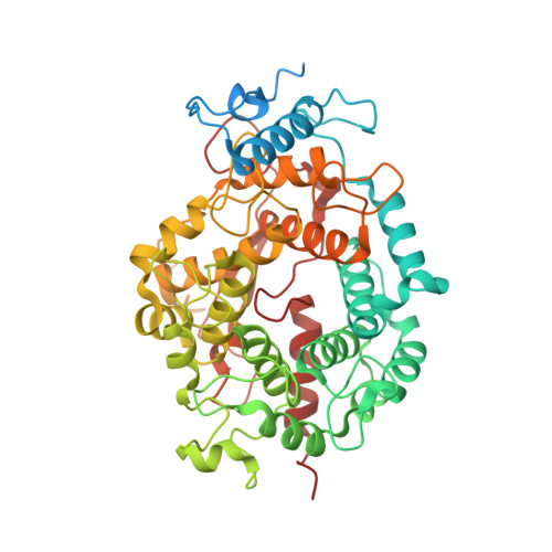
Crystal structures of the fungal pathogen Aspergillus fumigatus protein farnesyltransferase complexed with substrates and inhibitors reveal features for antifungal drug design.

Mabanglo, M.F.Hast, M.A.Lubock, N.B.Hellinga, H.W.Beese, L.S.

(2014) Protein Sci 23: 289-301

  • DOI: https://doi.org/10.1002/pro.2411
  • Primary Citation of Related Structures:  
    4L9P, 4LNB, 4LNG, 4MBG

  • PubMed Abstract: 

    Species of the fungal genus Aspergillus are significant human and agricultural pathogens that are often refractory to existing antifungal treatments. Protein farnesyltransferase (FTase), a critical enzyme in eukaryotes, is an attractive potential target for antifungal drug discovery. We report high-resolution structures of A. fumigatus FTase (AfFTase) in complex with substrates and inhibitors. Comparison of structures with farnesyldiphosphate (FPP) bound in the absence or presence of peptide substrate, corresponding to successive steps in ordered substrate binding, revealed that the second substrate-binding step is accompanied by motions of a loop in the catalytic site. Re-examination of other FTase structures showed that this motion is conserved. The substrate- and product-binding clefts in the AfFTase active site are wider than in human FTase (hFTase). Widening is a consequence of small shifts in the α-helices that comprise the majority of the FTase structure, which in turn arise from sequence variation in the hydrophobic core of the protein. These structural effects are key features that distinguish fungal FTases from hFTase. Their variation results in differences in steady-state enzyme kinetics and inhibitor interactions and presents opportunities for developing selective anti-fungal drugs by exploiting size differences in the active sites. We illustrate the latter by comparing the interaction of ED5 and Tipifarnib with hFTase and AfFTase. In AfFTase, the wider groove enables ED5 to bind in the presence of FPP, whereas in hFTase it binds only in the absence of substrate. Tipifarnib binds similarly to both enzymes but makes less extensive contacts in AfFTase with consequently weaker binding.


  • Organizational Affiliation

    Department of Biochemistry, Duke University Medical Center, Durham, North Carolina, 27710.


Macromolecules
Find similar proteins by:  (by identity cutoff)  |  3D Structure
Entity ID: 1
MoleculeChains Sequence LengthOrganismDetailsImage
CaaX farnesyltransferase alpha subunit Ram2367Aspergillus fumigatus Af293Mutation(s): 1 
Gene Names: AFUA_4G07800
EC: 2.5.1
UniProt
Find proteins for Q4WP27 (Aspergillus fumigatus (strain ATCC MYA-4609 / CBS 101355 / FGSC A1100 / Af293))
Explore Q4WP27 
Go to UniProtKB:  Q4WP27
Entity Groups  
Sequence Clusters30% Identity50% Identity70% Identity90% Identity95% Identity100% Identity
UniProt GroupQ4WP27
Sequence Annotations
Expand
  • Reference Sequence
Find similar proteins by:  (by identity cutoff)  |  3D Structure
Entity ID: 2
MoleculeChains Sequence LengthOrganismDetailsImage
CaaX farnesyltransferase beta subunit Ram1519Aspergillus fumigatus Af293Mutation(s): 0 
Gene Names: AFUA_4G10330
EC: 2.5.1.58
UniProt
Find proteins for Q4WPS9 (Aspergillus fumigatus (strain ATCC MYA-4609 / CBS 101355 / FGSC A1100 / Af293))
Explore Q4WPS9 
Go to UniProtKB:  Q4WPS9
Entity Groups  
Sequence Clusters30% Identity50% Identity70% Identity90% Identity95% Identity100% Identity
UniProt GroupQ4WPS9
Sequence Annotations
Expand
  • Reference Sequence

Find similar proteins by:  Sequence   |   3D Structure  

Entity ID: 3
MoleculeChains Sequence LengthOrganismDetailsImage
LYS-CYS-VAL-VAL-MET (CAAX peptide)5N/AMutation(s): 0 
Sequence Annotations
Expand
  • Reference Sequence
Small Molecules
Ligands 4 Unique
IDChains Name / Formula / InChI Key2D Diagram3D Interactions
FII
Query on FII

Download Ideal Coordinates CCD File 
Q [auth B][(3,7,11-TRIMETHYL-DODECA-2,6,10-TRIENYLOXYCARBAMOYL)-METHYL]-PHOSPHONIC ACID
C17 H30 N O5 P
JAOBYUCYSAOLHS-XGGJEREUSA-N
ZN
Query on ZN

Download Ideal Coordinates CCD File 
J [auth B]ZINC ION
Zn
PTFCDOFLOPIGGS-UHFFFAOYSA-N
EDO
Query on EDO

Download Ideal Coordinates CCD File 
E [auth A]
F [auth A]
G [auth A]
H [auth A]
I [auth A]
E [auth A],
F [auth A],
G [auth A],
H [auth A],
I [auth A],
M [auth B],
N [auth B],
O [auth B],
P [auth B]
1,2-ETHANEDIOL
C2 H6 O2
LYCAIKOWRPUZTN-UHFFFAOYSA-N
CL
Query on CL

Download Ideal Coordinates CCD File 
D [auth A],
K [auth B],
L [auth B]
CHLORIDE ION
Cl
VEXZGXHMUGYJMC-UHFFFAOYSA-M
Experimental Data & Validation

Experimental Data

  • Method: X-RAY DIFFRACTION
  • Resolution: 1.45 Å
  • R-Value Free: 0.152 
  • R-Value Work: 0.125 
  • R-Value Observed: 0.126 
  • Space Group: P 1 21 1
Unit Cell:
Length ( Å )Angle ( ˚ )
a = 63.236α = 90
b = 90.328β = 111.01
c = 83.007γ = 90
Software Package:
Software NamePurpose
ADSCdata collection
PHASESphasing
PHENIXrefinement
HKL-2000data reduction
HKL-2000data scaling

Structure Validation

View Full Validation Report


Ligand Structure Quality Assessment 


Entry History 

Deposition Data

Revision History  (Full details and data files)

  • Version 1.0: 2014-01-01
    Type: Initial release
  • Version 1.1: 2014-03-26
    Changes: Database references
  • Version 1.2: 2024-02-28
    Changes: Data collection, Database references, Derived calculations
  • Version 1.3: 2024-04-03
    Changes: Refinement description