4L68

Structure of the psedudokinase domain of BIR2, an immune regulator of the RLK/Pelle family


Experimental Data Snapshot

  • Method: X-RAY DIFFRACTION
  • Resolution: 2.00 Å
  • R-Value Free: 0.213 
  • R-Value Work: 0.175 
  • R-Value Observed: 0.177 

wwPDB Validation   3D Report Full Report


This is version 1.2 of the entry. See complete history


Literature

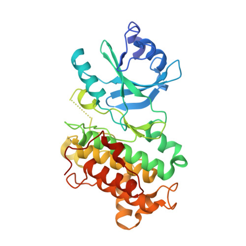
Structure of the pseudokinase domain of BIR2, a regulator of BAK1-mediated immune signaling in Arabidopsis.

Blaum, B.S.Mazzotta, S.Noldeke, E.R.Halter, T.Madlung, J.Kemmerling, B.Stehle, T.

(2014) J Struct Biol 186: 112-121

  • DOI: https://doi.org/10.1016/j.jsb.2014.02.005
  • Primary Citation of Related Structures:  
    4L68

  • PubMed Abstract: 

    The BAK1-interacting receptor-like kinase 2 (BIR2) belongs to the large family of leucine-rich repeat receptor-like kinases (LRR-RLKs) that mediate development and innate immunity in plants and form a monophyletic gene family with the Drosophila Pelle and human interleukin-1 receptor-associated kinases (IRAK). BIR2 is a negative regulator of BAK1-mediated defense mechanisms and cell death responses, yet key residues that are typically required for kinase activity are not present in the BIR2 kinase domain. We have determined the crystal structure of the BIR2 cytosolic domain and show that its nucleotide binding site is occluded. NMR spectroscopy confirmed that neither wild type nor phosphorylation-mimicking mutants of BIR2 bind ATP-analogues in solution, suggesting that BIR2 is a genuine enzymatically inactive pseudokinase. BIR2 is, however, phosphorylated by its target of regulation, BAK1. Using nano LC-MS/MS analysis for site-specific analysis of phosphorylation, we found a high density of BAK1-transphosphorylation sites in the BIR2 juxta membrane domain, a region previously implicated in regulation of RLKs. Our findings provide a structural basis to better understand signaling through kinase-dead domains that are predicted to account for 20% of all Arabidopsis RLKs and 10% of all human kinases.


  • Organizational Affiliation

    Interfaculty Institute of Biochemistry, University of Tübingen, 72076 Tübingen, Germany.


Macromolecules
Find similar proteins by:  (by identity cutoff)  |  3D Structure
Entity ID: 1
MoleculeChains Sequence LengthOrganismDetailsImage
Leucine-rich repeat protein kinase-like protein
A, B
340Arabidopsis thalianaMutation(s): 0 
Gene Names: AT3G28450At3g28450/MFJ20_13LRR-RLK
UniProt
Find proteins for Q9LSI9 (Arabidopsis thaliana)
Explore Q9LSI9 
Go to UniProtKB:  Q9LSI9
Entity Groups  
Sequence Clusters30% Identity50% Identity70% Identity90% Identity95% Identity100% Identity
UniProt GroupQ9LSI9
Sequence Annotations
Expand
  • Reference Sequence
Small Molecules
Ligands 1 Unique
IDChains Name / Formula / InChI Key2D Diagram3D Interactions
PDO
Query on PDO

Download Ideal Coordinates CCD File 
C [auth A]1,3-PROPANDIOL
C3 H8 O2
YPFDHNVEDLHUCE-UHFFFAOYSA-N
Experimental Data & Validation

Experimental Data

  • Method: X-RAY DIFFRACTION
  • Resolution: 2.00 Å
  • R-Value Free: 0.213 
  • R-Value Work: 0.175 
  • R-Value Observed: 0.177 
  • Space Group: H 3 2
Unit Cell:
Length ( Å )Angle ( ˚ )
a = 108.141α = 90
b = 108.141β = 90
c = 392.655γ = 120
Software Package:
Software NamePurpose
TVXdata collection
CCP4model building
PHENIXrefinement
XDSdata reduction
XDSdata scaling
CCP4phasing

Structure Validation

View Full Validation Report


Entry History 

Deposition Data

Revision History  (Full details and data files)

  • Version 1.0: 2014-03-05
    Type: Initial release
  • Version 1.1: 2014-04-23
    Changes: Database references
  • Version 1.2: 2024-02-28
    Changes: Data collection, Database references, Derived calculations