4KW6

Crystal structure of Peroxiredoxin-1 (C-terminal truncation mutant) from the human hookworm Ancylostoma ceylanicum bound to conoidin A


Experimental Data Snapshot

  • Method: X-RAY DIFFRACTION
  • Resolution: 3.00 Å
  • R-Value Free: 0.200 
  • R-Value Work: 0.186 
  • R-Value Observed: 0.187 

wwPDB Validation   3D Report Full Report


Ligand Structure Quality Assessment 


This is version 1.3 of the entry. See complete history


Literature

Peroxiredoxin-1 from the Human Hookworm Ancylostoma ceylanicum Forms a Stable Oxidized Decamer and Is Covalently Inhibited by Conoidin A.

Nguyen, J.B.Pool, C.D.Wong, C.Y.Treger, R.S.Williams, D.L.Cappello, M.Lea, W.A.Simeonov, A.Vermeire, J.J.Modis, Y.

(2013) Chem Biol 20: 991-1001

  • DOI: https://doi.org/10.1016/j.chembiol.2013.06.011
  • Primary Citation of Related Structures:  
    4FH8, 4KW6

  • PubMed Abstract: 

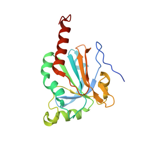
    Hookworms are parasitic nematodes that have a devastating impact on global health, particularly in developing countries. We report a biochemical and structural analysis of a peroxiredoxin from the hookworm Ancylostoma ceylanicum, AcePrx-1. Peroxiredoxins provide antioxidant protection and act as signaling molecules and chaperones. AcePrx-1 is expressed in adult hookworms and can be inactivated by 2,3-bis(bromomethyl)quinoxaline-1,4-dioxide (conoidin A). Conoidin A inactivates AcePrx-1 by alkylating or crosslinking the catalytic cysteines, while maintaining the enzyme in the "locally unfolded" conformation. Irreversible oxidation of the resolving cysteine may contribute additional inhibitory activity. A crystal structure of oxidized AcePrx-1 reveals a disulfide-linked decamer. A helix macrodipole near the active site increases the reactivity of the catalytic cysteines to conoidin A. This work demonstrates the promise of conoidin compounds as probes to evaluate peroxiredoxins as drug targets in human parasites.


  • Organizational Affiliation

    Department of Molecular Biophysics and Biochemistry, Yale University, New Haven, CT 06520, USA.


Macromolecules
Find similar proteins by:  (by identity cutoff)  |  3D Structure
Entity ID: 1
MoleculeChains Sequence LengthOrganismDetailsImage
Peroxiredoxin-1
A, B, C, D, E
179Ancylostoma ceylanicumMutation(s): 0 
EC: 1.11.1.15
UniProt
Find proteins for J7HJM3 (Ancylostoma ceylanicum)
Explore J7HJM3 
Go to UniProtKB:  J7HJM3
Entity Groups  
Sequence Clusters30% Identity50% Identity70% Identity90% Identity95% Identity100% Identity
UniProt GroupJ7HJM3
Sequence Annotations
Expand
  • Reference Sequence
Small Molecules
Ligands 1 Unique
IDChains Name / Formula / InChI Key2D Diagram3D Interactions
QDO
Query on QDO

Download Ideal Coordinates CCD File 
F [auth B],
G [auth C]
2,3-bis(bromomethyl)quinoxaline 1,4-dioxide
C10 H8 Br2 N2 O2
DQKNFTLRMZOAMG-UHFFFAOYSA-N
Binding Affinity Annotations 
IDSourceBinding Affinity
QDO PDBBind:  4KW6 IC50: 3.74e+5 (nM) from 1 assay(s)
Experimental Data & Validation

Experimental Data

  • Method: X-RAY DIFFRACTION
  • Resolution: 3.00 Å
  • R-Value Free: 0.200 
  • R-Value Work: 0.186 
  • R-Value Observed: 0.187 
  • Space Group: C 1 2 1
Unit Cell:
Length ( Å )Angle ( ˚ )
a = 141.554α = 90
b = 141.657β = 98.5
c = 68.855γ = 90
Software Package:
Software NamePurpose
HKL-2000data collection
PHASERphasing
REFMACrefinement
HKL-2000data reduction
HKL-2000data scaling

Structure Validation

View Full Validation Report


Ligand Structure Quality Assessment 


Entry History 

Deposition Data

Revision History  (Full details and data files)

  • Version 1.0: 2013-07-31
    Type: Initial release
  • Version 1.1: 2013-08-14
    Changes: Database references
  • Version 1.2: 2013-09-11
    Changes: Database references
  • Version 1.3: 2023-09-20
    Changes: Data collection, Database references, Derived calculations, Refinement description