4IIT

The Phenylacetyl-CoA monooxygenase PaaABC subcomplex with phenylacetyl-CoA


Experimental Data Snapshot

  • Method: X-RAY DIFFRACTION
  • Resolution: 4.30 Å
  • R-Value Free: 0.363 
  • R-Value Work: 0.288 
  • R-Value Observed: 0.291 

wwPDB Validation   3D Report Full Report


Ligand Structure Quality Assessment 


This is version 1.5 of the entry. See complete history


Literature

Family of phenylacetyl-CoA monooxygenases differs in subunit organization from other monooxygenases.

Grishin, A.M.Ajamian, E.Tao, L.Bostina, M.Zhang, L.Trempe, J.F.Menard, R.Rouiller, I.Cygler, M.

(2013) J Struct Biol 184: 147-154

  • DOI: https://doi.org/10.1016/j.jsb.2013.09.012
  • Primary Citation of Related Structures:  
    4IIT

  • PubMed Abstract: 

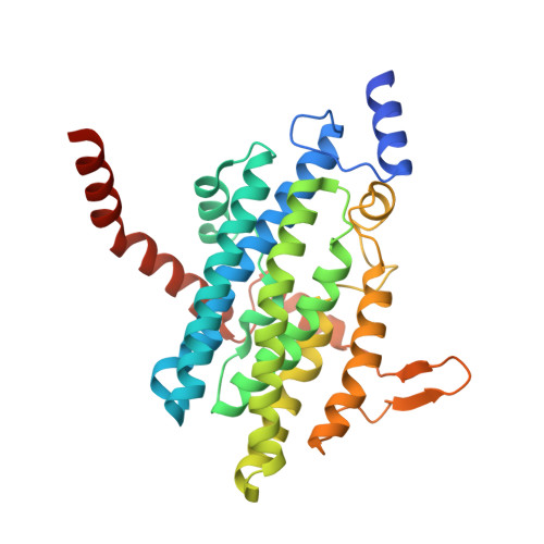
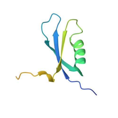
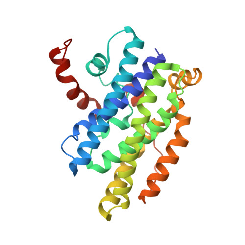
    The phenylacetate degradation pathway is present in a wide range of microbes. A key component of this pathway is the four-subunit phenylacetyl-coenzyme A monooxygenase complex (PA-CoA MO, PaaACBE) that catalyzes the insertion of an oxygen in the aromatic ring of PA. This multicomponent enzyme represents a new family of monooxygenases. We have previously determined the structure of the PaaAC subcomplex of catalytic (A) and structural (C) subunits and shown that PaaACB form a stable complex. The PaaB subunit is unrelated to the small subunits of homologous monooxygenases and its role and organization of the PaaACB complex is unknown. From low-resolution crystal structure, electron microscopy and small angle X-ray scattering we show that the PaaACB complex forms heterohexamers, with a homodimer of PaaB bridging two PaaAC heterodimers. Modeling the interactions of reductase subunit PaaE with PaaACB suggested that a unique and conserved 'lysine bridge' constellation near the Fe-binding site in the PaaA subunit (Lys68, Glu49, Glu72 and Asp126) may form part of the electron transfer path from PaaE to the iron center. The crystal structure of the PaaA(K68Q/E49Q)-PaaC is very similar to the wild-type enzyme structure, but when combined with the PaaE subunit the mutant showed 20-50 times reduced activity, supporting the functional importance of the 'lysine bridge'.


  • Organizational Affiliation

    Department of Biochemistry, University of Saskatchewan, Saskatoon, Saskatchewan S7N 5E5, Canada.


Macromolecules
Find similar proteins by:  (by identity cutoff)  |  3D Structure
Entity ID: 1
MoleculeChains Sequence LengthOrganismDetailsImage
Phenylacetate-CoA oxygenase subunit PaaA320Klebsiella pneumoniae subsp. pneumoniae MGH 78578Mutation(s): 0 
Gene Names: KPHS_23690paaA
EC: 1.14.13
UniProt
Find proteins for A6T8I0 (Klebsiella pneumoniae subsp. pneumoniae (strain ATCC 700721 / MGH 78578))
Explore A6T8I0 
Go to UniProtKB:  A6T8I0
Entity Groups  
Sequence Clusters30% Identity50% Identity70% Identity90% Identity95% Identity100% Identity
UniProt GroupA6T8I0
Sequence Annotations
Expand
  • Reference Sequence
Find similar proteins by:  (by identity cutoff)  |  3D Structure
Entity ID: 2
MoleculeChains Sequence LengthOrganismDetailsImage
Phenylacetate-CoA oxygenase subunit PaaB95Klebsiella pneumoniae subsp. pneumoniae MGH 78578Mutation(s): 0 
Gene Names: EAE_20515paaB
EC: 1.14.13
UniProt
Find proteins for A6T8I1 (Klebsiella pneumoniae subsp. pneumoniae (strain ATCC 700721 / MGH 78578))
Explore A6T8I1 
Go to UniProtKB:  A6T8I1
Entity Groups  
Sequence Clusters30% Identity50% Identity70% Identity90% Identity95% Identity100% Identity
UniProt GroupA6T8I1
Sequence Annotations
Expand
  • Reference Sequence
Find similar proteins by:  (by identity cutoff)  |  3D Structure
Entity ID: 3
MoleculeChains Sequence LengthOrganismDetailsImage
Phenylacetate-CoA oxygenase subunit PaaC251Klebsiella pneumoniae subsp. pneumoniae MGH 78578Mutation(s): 0 
Gene Names: KPN78578_14420KPN_01471paaC
EC: 1.14.13
UniProt
Find proteins for A6T8I2 (Klebsiella pneumoniae subsp. pneumoniae (strain ATCC 700721 / MGH 78578))
Explore A6T8I2 
Go to UniProtKB:  A6T8I2
Entity Groups  
Sequence Clusters30% Identity50% Identity70% Identity90% Identity95% Identity100% Identity
UniProt GroupA6T8I2
Sequence Annotations
Expand
  • Reference Sequence
Small Molecules
Ligands 1 Unique
IDChains Name / Formula / InChI Key2D Diagram3D Interactions
FAQ
Query on FAQ

Download Ideal Coordinates CCD File 
D [auth A]Phenylacetyl coenzyme A
C29 H42 N7 O17 P3 S
ZIGIFDRJFZYEEQ-CECATXLMSA-N
Experimental Data & Validation

Experimental Data

  • Method: X-RAY DIFFRACTION
  • Resolution: 4.30 Å
  • R-Value Free: 0.363 
  • R-Value Work: 0.288 
  • R-Value Observed: 0.291 
  • Space Group: P 63 2 2
Unit Cell:
Length ( Å )Angle ( ˚ )
a = 208.941α = 90
b = 208.941β = 90
c = 86.081γ = 120
Software Package:
Software NamePurpose
PHENIXrefinement
PHASERphasing
REFMACrefinement
PDB_EXTRACTdata extraction
XDSdata scaling
DENZOdata reduction
HKL-2000data scaling
SCALEPACKdata scaling

Structure Validation

View Full Validation Report


Ligand Structure Quality Assessment 


Entry History 

Deposition Data

Revision History  (Full details and data files)

  • Version 1.0: 2013-11-06
    Type: Initial release
  • Version 1.1: 2013-12-25
    Changes: Database references
  • Version 1.2: 2017-11-15
    Changes: Refinement description
  • Version 1.3: 2018-01-24
    Changes: Refinement description
  • Version 1.4: 2019-07-17
    Changes: Data collection, Refinement description
  • Version 1.5: 2023-09-20
    Changes: Data collection, Database references, Derived calculations, Refinement description