4IDW

Polycrystalline T6 Bovine Insulin: Anisotropic Lattice Evolution and Novel Structure Refinement Strategy


Experimental Data Snapshot

  • Method: POWDER DIFFRACTION

wwPDB Validation   3D Report Full Report


This is version 1.0 of the entry. See complete history


Literature

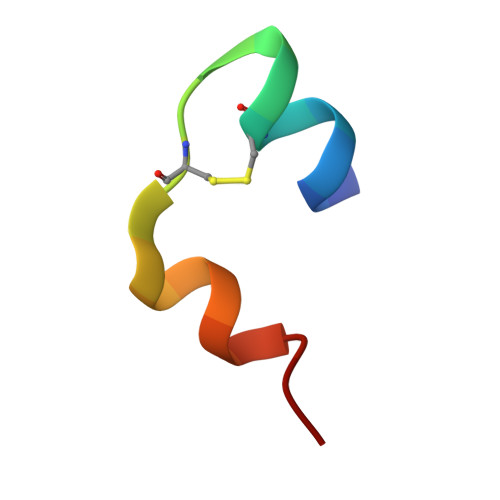
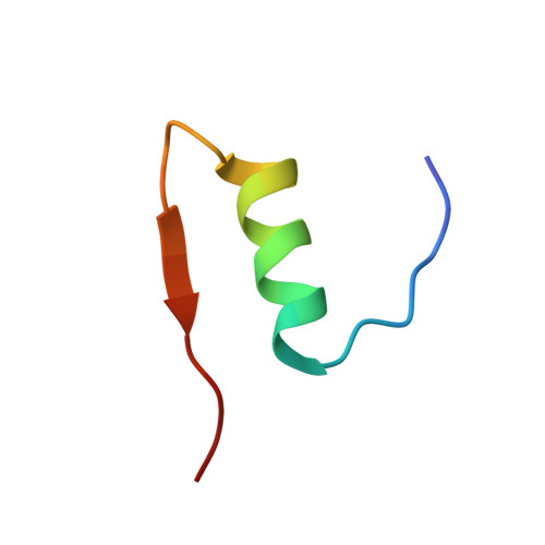
High-resolution powder X-ray data reveal the T6 hexameric form of bovine insulin

Margiolaki, I.Giannopoulou, A.E.Wright, J.P.Knight, L.Norrman, M.Schluckebier, G.Fitch, A.N.Von Dreele, R.B.

(2013) Acta Crystallogr D Biol Crystallogr 69: 978-990

  • DOI: https://doi.org/10.1107/S0907444913003867
  • Primary Citation of Related Structures:  
    4IDW

  • PubMed Abstract: 

    A series of bovine insulin samples were obtained as 14 polycrystalline precipitates at room temperature in the pH range 5.0-7.6. High-resolution powder X-ray diffraction data were collected to reveal the T6 hexameric insulin form. Sample homogeneity and reproducibility were verified by additional synchrotron measurements using an area detector. Pawley analyses of the powder patterns displayed pH- and radiation-induced anisotropic lattice modifications. The pronounced anisotropic lattice variations observed for T6 insulin were exploited in a 14-data-set Rietveld refinement to obtain an average crystal structure over the pH range investigated. Only the protein atoms of the known structure with PDB code 2a3g were employed in our starting model. A novel approach for refining protein structures using powder diffraction data is presented. In this approach, each amino acid is represented by a flexible rigid body (FRB). The FRB model requires a significantly smaller number of refinable parameters and restraints than a fully free-atom refinement. A total of 1542 stereochemical restraints were imposed in order to refine the positions of 800 protein atoms, two Zn atoms and 44 water molecules in the asymmetric unit using experimental data in the resolution range 18.2-2.7 Å for all profiles.


  • Organizational Affiliation

    Department of Biology, Section of Genetics, Cell Biology and Development, University of Patras, GR-26500 Patras, Greece. imargiola@upatras.gr


Macromolecules

Find similar proteins by:  Sequence   |   3D Structure  

Entity ID: 1
MoleculeChains Sequence LengthOrganismDetailsImage
Insulin A chain
A, C
21Bos taurusMutation(s): 0 
UniProt
Find proteins for P01317 (Bos taurus)
Explore P01317 
Go to UniProtKB:  P01317
Entity Groups  
Sequence Clusters30% Identity50% Identity70% Identity90% Identity95% Identity100% Identity
UniProt GroupP01317
Sequence Annotations
Expand
  • Reference Sequence
Find similar proteins by:  (by identity cutoff)  |  3D Structure
Entity ID: 2
MoleculeChains Sequence LengthOrganismDetailsImage
Insulin B chain
B, D
30Bos taurusMutation(s): 0 
UniProt
Find proteins for P01317 (Bos taurus)
Explore P01317 
Go to UniProtKB:  P01317
Entity Groups  
Sequence Clusters30% Identity50% Identity70% Identity90% Identity95% Identity100% Identity
UniProt GroupP01317
Sequence Annotations
Expand
  • Reference Sequence
Small Molecules
Ligands 1 Unique
IDChains Name / Formula / InChI Key2D Diagram3D Interactions
ZN
Query on ZN

Download Ideal Coordinates CCD File 
E [auth B],
F [auth D]
ZINC ION
Zn
PTFCDOFLOPIGGS-UHFFFAOYSA-N
Experimental Data & Validation

Experimental Data

  • Method: POWDER DIFFRACTION
  • Space Group: H 3
Unit Cell:
Length ( Å )Angle ( ˚ )
a = 82.607α = 90
b = 82.607β = 90
c = 33.632γ = 120
Software Package:
Software NamePurpose
ID31sumdata collection
MOLREPphasing
GSASrefinement
FIT2Ddata reduction
ID31sumdata scaling

Structure Validation

View Full Validation Report


Entry History 

Deposition Data

Revision History  (Full details and data files)

  • Version 1.0: 2013-06-05
    Type: Initial release