4IA5

Hydratase from Lactobacillus acidophilus - SeMet derivative (apo LAH)


Experimental Data Snapshot

  • Method: X-RAY DIFFRACTION
  • Resolution: 2.22 Å
  • R-Value Free: 0.259 
  • R-Value Work: 0.214 
  • R-Value Observed: 0.216 

wwPDB Validation   3D Report Full Report


This is version 1.1 of the entry. See complete history


Literature

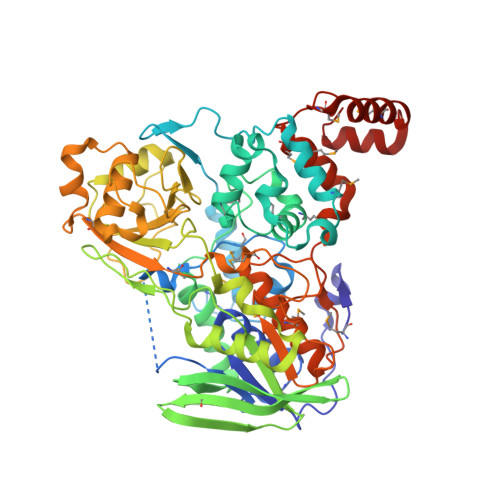
Crystal structure analysis of a fatty acid double-bond hydratase from Lactobacillus acidophilus

Volkov, A.Khoshnevis, S.Neumann, P.Herrfurth, C.Wohlwend, D.Ficner, R.Feussner, I.

(2013) Acta Crystallogr D Biol Crystallogr 69: 648-657

  • DOI: https://doi.org/10.1107/S0907444913000991
  • Primary Citation of Related Structures:  
    4IA5, 4IA6

  • PubMed Abstract: 

    Bacteria have evolved mechanisms for the hydrogenation of unsaturated fatty acids. Hydroxy fatty acid formation may be the first step in such a process; however, knowledge of the structural and mechanistic aspects of this reaction is scarce. Recently, myosin cross-reactive antigen was shown to be a bacterial FAD-containing hydratase which acts on the 9Z and 12Z double bonds of C16 and C18 non-esterified fatty acids, with the formation of 10-hydroxy and 10,13-dihydroxy fatty acids. These fatty acid hydratases form a large protein family which is conserved across Gram-positive and Gram-negative bacteria with no sequence similarity to any known protein apart from the FAD-binding motif. In order to shed light on the substrate recognition and the mechanism of the hydratase reaction, the crystal structure of the hydratase from Lactobacillus acidophilus (LAH) was determined by single-wavelength anomalous dispersion. Crystal structures of apo LAH and of LAH with bound linoleic acid were refined at resolutions of 2.3 and 1.8 Å, respectively. LAH is a homodimer; each protomer consists of four intricately connected domains. Three of them form the FAD-binding and substrate-binding sites and reveal structural similarity to three domains of several flavin-dependent enzymes, including amine oxidoreductases. The additional fourth domain of LAH is located at the C-terminus and consists of three α-helices. It covers the entrance to the hydrophobic substrate channel leading from the protein surface to the active site. In the presence of linoleic acid, the fourth domain of one protomer undergoes conformational changes and opens the entrance to the substrate-binding channel of the other protomer of the LAH homodimer. The linoleic acid molecule is bound at the entrance to the substrate channel, suggesting movement of the lid domain triggered by substrate recognition.


  • Organizational Affiliation

    Department for Plant Biochemistry, Albrecht-von-Haller-Institute for Plant Sciences, Georg-August-University, Göttingen, Germany.


Macromolecules
Find similar proteins by:  (by identity cutoff)  |  3D Structure
Entity ID: 1
MoleculeChains Sequence LengthOrganismDetailsImage
Myosin-crossreactive antigen
A, B
591Lactobacillus acidophilus NCFMMutation(s): 0 
Gene Names: LBA0649
UniProt
Find proteins for Q5FL96 (Lactobacillus acidophilus (strain ATCC 700396 / NCK56 / N2 / NCFM))
Explore Q5FL96 
Go to UniProtKB:  Q5FL96
Entity Groups  
Sequence Clusters30% Identity50% Identity70% Identity90% Identity95% Identity100% Identity
UniProt GroupQ5FL96
Sequence Annotations
Expand
  • Reference Sequence
Small Molecules
Ligands 6 Unique
IDChains Name / Formula / InChI Key2D Diagram3D Interactions
MES
Query on MES

Download Ideal Coordinates CCD File 
C [auth A],
J [auth B]
2-(N-MORPHOLINO)-ETHANESULFONIC ACID
C6 H13 N O4 S
SXGZJKUKBWWHRA-UHFFFAOYSA-N
MPD
Query on MPD

Download Ideal Coordinates CCD File 
F [auth A],
G [auth A],
H [auth A],
L [auth B],
M [auth B]
(4S)-2-METHYL-2,4-PENTANEDIOL
C6 H14 O2
SVTBMSDMJJWYQN-YFKPBYRVSA-N
PO4
Query on PO4

Download Ideal Coordinates CCD File 
K [auth B]PHOSPHATE ION
O4 P
NBIIXXVUZAFLBC-UHFFFAOYSA-K
GOL
Query on GOL

Download Ideal Coordinates CCD File 
N [auth B],
O [auth B],
P [auth B]
GLYCEROL
C3 H8 O3
PEDCQBHIVMGVHV-UHFFFAOYSA-N
K
Query on K

Download Ideal Coordinates CCD File 
D [auth A],
E [auth A]
POTASSIUM ION
K
NPYPAHLBTDXSSS-UHFFFAOYSA-N
NA
Query on NA

Download Ideal Coordinates CCD File 
I [auth B]SODIUM ION
Na
FKNQFGJONOIPTF-UHFFFAOYSA-N
Modified Residues  1 Unique
IDChains TypeFormula2D DiagramParent
MSE
Query on MSE
A, B
L-PEPTIDE LINKINGC5 H11 N O2 SeMET
Experimental Data & Validation

Experimental Data

  • Method: X-RAY DIFFRACTION
  • Resolution: 2.22 Å
  • R-Value Free: 0.259 
  • R-Value Work: 0.214 
  • R-Value Observed: 0.216 
  • Space Group: P 21 21 2
Unit Cell:
Length ( Å )Angle ( ˚ )
a = 168.75α = 90
b = 78.97β = 90
c = 108.77γ = 90
Software Package:
Software NamePurpose
XSCALEdata scaling
SHELXphasing
PHENIXrefinement
PDB_EXTRACTdata extraction
DNAdata collection
XDSdata reduction
SHELXDphasing

Structure Validation

View Full Validation Report


Entry History 

Deposition Data

Revision History  (Full details and data files)

  • Version 1.0: 2013-03-27
    Type: Initial release
  • Version 1.1: 2013-08-07
    Changes: Database references