4H2B

Human ecto-5'-nucleotidase (CD73): crystal form II (open) in complex with Baicalin


Experimental Data Snapshot

  • Method: X-RAY DIFFRACTION
  • Resolution: 1.70 Å
  • R-Value Free: 0.180 
  • R-Value Work: 0.150 
  • R-Value Observed: 0.151 

wwPDB Validation   3D Report Full Report


Ligand Structure Quality Assessment 


This is version 1.2 of the entry. See complete history


Literature

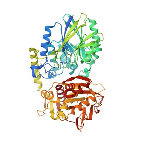
Crystal Structure of the Human Ecto-5'-Nucleotidase (CD73): Insights into the Regulation of Purinergic Signaling.

Knapp, K.Zebisch, M.Pippel, J.El-Tayeb, A.Muller, C.E.Strater, N.

(2012) Structure 20: 2161-2173

  • DOI: https://doi.org/10.1016/j.str.2012.10.001
  • Primary Citation of Related Structures:  
    4H1Y, 4H2B, 4H2F, 4H2G, 4H2I

  • PubMed Abstract: 

    In vertebrates ecto-5'-nucleotidase (e5NT) catalyzes the hydrolysis of extracellular AMP to adenosine and represents the major control point for extracellular adenosine levels. Due to its pivotal role for activation of P1 adenosine receptors, e5NT has emerged as an appealing drug target for treatment of inflammation, chronic pain, hypoxia, and cancer. Crystal structures of the dimeric human e5NT reveal an extensive 114° conformational switch between the open and closed forms of the enzyme. The dimerization interface is formed by the C-terminal domains and exhibits interchain motions of up to 13°. Complex structures with adenosine and AMPCP indicate that structural control of the domain movement determines the selectivity for monophosphate nucleotides. Binding modes of nucleotide-derived and flavonoid-based compounds complexed to the C-terminal domain in the open form reveal an additional binding pocket of ∼210 Å(3) that might be explored to design more potent inhibitors.


  • Organizational Affiliation

    Institute of Bioanalytical Chemistry, Center for Biotechnology and Biomedicine, University of Leipzig, Deutscher Platz 5, 04103 Leipzig, Germany.


Macromolecules
Find similar proteins by:  (by identity cutoff)  |  3D Structure
Entity ID: 1
MoleculeChains Sequence LengthOrganismDetailsImage
5'-nucleotidase547Homo sapiensMutation(s): 8 
Gene Names: NT5NT5ENTE
EC: 3.1.3.5
UniProt & NIH Common Fund Data Resources
Find proteins for P21589 (Homo sapiens)
Explore P21589 
Go to UniProtKB:  P21589
PHAROS:  P21589
GTEx:  ENSG00000135318 
Entity Groups  
Sequence Clusters30% Identity50% Identity70% Identity90% Identity95% Identity100% Identity
UniProt GroupP21589
Sequence Annotations
Expand
  • Reference Sequence
Experimental Data & Validation

Experimental Data

  • Method: X-RAY DIFFRACTION
  • Resolution: 1.70 Å
  • R-Value Free: 0.180 
  • R-Value Work: 0.150 
  • R-Value Observed: 0.151 
  • Space Group: P 21 21 2
Unit Cell:
Length ( Å )Angle ( ˚ )
a = 67.263α = 90
b = 131.056β = 90
c = 66.238γ = 90
Software Package:
Software NamePurpose
REFMACrefinement

Structure Validation

View Full Validation Report


Ligand Structure Quality Assessment 


Entry History 

Deposition Data

Revision History  (Full details and data files)

  • Version 1.0: 2012-11-28
    Type: Initial release
  • Version 1.1: 2012-12-12
    Changes: Structure summary
  • Version 1.2: 2012-12-26
    Changes: Database references