4GR8

Crystal structure of the catalytic domain of Human MMP12 in complex with selective phosphinic inhibitor RXP470C


Experimental Data Snapshot

  • Method: X-RAY DIFFRACTION
  • Resolution: 1.30 Å
  • R-Value Free: 0.203 
  • R-Value Work: 0.177 
  • R-Value Observed: 0.178 

wwPDB Validation   3D Report Full Report


Ligand Structure Quality Assessment 


This is version 1.2 of the entry. See complete history


Literature

Molecular determinants of a selective matrix metalloprotease-12 inhibitor: insights from crystallography and thermodynamic studies.

Czarny, B.Stura, E.A.Devel, L.Vera, L.Cassar-Lajeunesse, E.Beau, F.Calderone, V.Fragai, M.Luchinat, C.Dive, V.

(2013) J Med Chem 56: 1149-1159

  • DOI: https://doi.org/10.1021/jm301574d
  • Primary Citation of Related Structures:  
    4GQL, 4GR0, 4GR3, 4GR8

  • PubMed Abstract: 

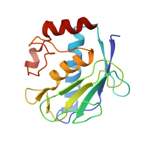
    The molecular determinants responsible for the potency of the RXP470.1 phosphinic peptide inhibitor toward matrix metalloprotease-12 (MMP-12) remain elusive. To address this issue, structure-activity study, X-ray crystallography, and isothermal titration calorimetry (ITC) experiments were performed. The crystal structure of MMP-12/inhibitor complex (1.15 Å) reveals that the inhibitor establishes multiple interactions with the MMP-12 active site, with its long P(1)' side chain filling most of the S(1)' deep cavity. ITC experiments indicate that the binding of this inhibitor to MMP-12 is mostly entropy driven (ΔG° = -13.1 kcal/mol, ΔH° = -2.53 kcal/mol, and -TΔS° = -10.60 kcal/mol) and involves a proton uptake from the buffer. Comparing phosphinic versus hydroxamate inhibitors reveals that the chelation of the zinc ion is slightly different, leading the inhibitor backbone to adopt a position in which the hydrogen bonding with the MMP-12 active site is less favorable in phosphinic inhibitor while maintaining high affinity.


  • Organizational Affiliation

    CEA, Service d'Ingénierie Moléculaire des Protéines (SIMOPRO), Gif/Yvette 91191 Cedex, France.


Macromolecules
Find similar proteins by:  (by identity cutoff)  |  3D Structure
Entity ID: 1
MoleculeChains Sequence LengthOrganismDetailsImage
Macrophage metalloelastase152Homo sapiensMutation(s): 1 
Gene Names: HMEMMP12
EC: 3.4.24.65
UniProt & NIH Common Fund Data Resources
Find proteins for P39900 (Homo sapiens)
Explore P39900 
Go to UniProtKB:  P39900
PHAROS:  P39900
GTEx:  ENSG00000262406 
Entity Groups  
Sequence Clusters30% Identity50% Identity70% Identity90% Identity95% Identity100% Identity
UniProt GroupP39900
Sequence Annotations
Expand
  • Reference Sequence
Small Molecules
Ligands 4 Unique
IDChains Name / Formula / InChI Key2D Diagram3D Interactions
R4C
Query on R4C

Download Ideal Coordinates CCD File 
G [auth A]N-{(2S)-3-[(R)-(4-bromophenyl)(hydroxy)phosphoryl]-2-[(3-phenyl-1,2-oxazol-5-yl)methyl]propanoyl}-L-alanyl-L-alaninamid e
C25 H28 Br N4 O6 P
HXHLWPVBBLIGJV-XYJFISCASA-N
IMD
Query on IMD

Download Ideal Coordinates CCD File 
H [auth A]IMIDAZOLE
C3 H5 N2
RAXXELZNTBOGNW-UHFFFAOYSA-O
ZN
Query on ZN

Download Ideal Coordinates CCD File 
B [auth A],
C [auth A]
ZINC ION
Zn
PTFCDOFLOPIGGS-UHFFFAOYSA-N
CA
Query on CA

Download Ideal Coordinates CCD File 
D [auth A],
E [auth A],
F [auth A]
CALCIUM ION
Ca
BHPQYMZQTOCNFJ-UHFFFAOYSA-N
Binding Affinity Annotations 
IDSourceBinding Affinity
R4C PDBBind:  4GR8 Ki: 14.7 (nM) from 1 assay(s)
Binding MOAD:  4GR8 Ki: 14.7 (nM) from 1 assay(s)
BindingDB:  4GR8 Ki: 15 (nM) from 1 assay(s)
Experimental Data & Validation

Experimental Data

  • Method: X-RAY DIFFRACTION
  • Resolution: 1.30 Å
  • R-Value Free: 0.203 
  • R-Value Work: 0.177 
  • R-Value Observed: 0.178 
  • Space Group: P 21 21 2
Unit Cell:
Length ( Å )Angle ( ˚ )
a = 67.83α = 90
b = 61.47β = 90
c = 33.46γ = 90
Software Package:
Software NamePurpose
CBASSdata collection
MOLREPphasing
PHENIXrefinement
XDSdata reduction
XDSdata scaling

Structure Validation

View Full Validation Report


Ligand Structure Quality Assessment 


Entry History 

Deposition Data

Revision History  (Full details and data files)

  • Version 1.0: 2013-02-06
    Type: Initial release
  • Version 1.1: 2013-05-22
    Changes: Database references
  • Version 1.2: 2023-09-13
    Changes: Data collection, Database references, Derived calculations, Refinement description, Structure summary