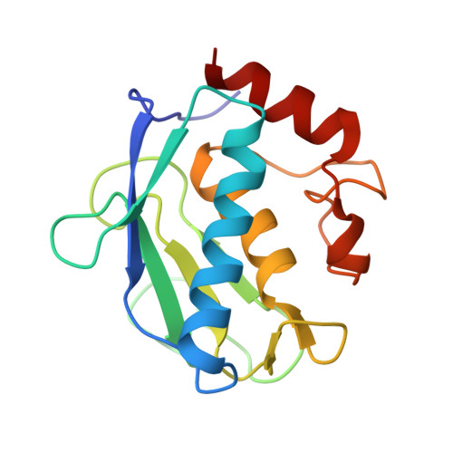
4GR3

Crystal structure of the catalytic domain of Human MMP12 in complex with selective phosphinic inhibitor RXP470A


Experimental Data Snapshot

  • Method: X-RAY DIFFRACTION
  • Resolution: 1.49 Å
  • R-Value Free: 0.216 
  • R-Value Work: 0.180 
  • R-Value Observed: 0.182 

wwPDB Validation   3D Report Full Report


This is version 1.2 of the entry. See complete history


Literature

Molecular determinants of a selective matrix metalloprotease-12 inhibitor: insights from crystallography and thermodynamic studies.

Czarny, B.Stura, E.A.Devel, L.Vera, L.Cassar-Lajeunesse, E.Beau, F.Calderone, V.Fragai, M.Luchinat, C.Dive, V.

(2013) J Med Chem 56: 1149-1159

  • DOI: https://doi.org/10.1021/jm301574d
  • Primary Citation of Related Structures:  
    4GQL, 4GR0, 4GR3, 4GR8

  • PubMed Abstract: 

    The molecular determinants responsible for the potency of the RXP470.1 phosphinic peptide inhibitor toward matrix metalloprotease-12 (MMP-12) remain elusive. To address this issue, structure-activity study, X-ray crystallography, and isothermal titration calorimetry (ITC) experiments were performed. The crystal structure of MMP-12/inhibitor complex (1.15 Å) reveals that the inhibitor establishes multiple interactions with the MMP-12 active site, with its long P(1)' side chain filling most of the S(1)' deep cavity. ITC experiments indicate that the binding of this inhibitor to MMP-12 is mostly entropy driven (ΔG° = -13.1 kcal/mol, ΔH° = -2.53 kcal/mol, and -TΔS° = -10.60 kcal/mol) and involves a proton uptake from the buffer. Comparing phosphinic versus hydroxamate inhibitors reveals that the chelation of the zinc ion is slightly different, leading the inhibitor backbone to adopt a position in which the hydrogen bonding with the MMP-12 active site is less favorable in phosphinic inhibitor while maintaining high affinity.


  • Organizational Affiliation

    CEA, Service d'Ingénierie Moléculaire des Protéines (SIMOPRO), Gif/Yvette 91191 Cedex, France.


Macromolecules
Find similar proteins by:  (by identity cutoff)  |  3D Structure
Entity ID: 1
MoleculeChains Sequence LengthOrganismDetailsImage
Macrophage metalloelastase159Homo sapiensMutation(s): 1 
Gene Names: HMEMMP12
EC: 3.4.24.65
UniProt & NIH Common Fund Data Resources
Find proteins for P39900 (Homo sapiens)
Explore P39900 
Go to UniProtKB:  P39900
PHAROS:  P39900
GTEx:  ENSG00000262406 
Entity Groups  
Sequence Clusters30% Identity50% Identity70% Identity90% Identity95% Identity100% Identity
UniProt GroupP39900
Sequence Annotations
Expand
  • Reference Sequence
Small Molecules
Binding Affinity Annotations 
IDSourceBinding Affinity
R45 BindingDB:  4GR3 Ki: 16 (nM) from 1 assay(s)
Binding MOAD:  4GR3 Ki: 15.5 (nM) from 1 assay(s)
PDBBind:  4GR3 Ki: 15.5 (nM) from 1 assay(s)
Experimental Data & Validation

Experimental Data

  • Method: X-RAY DIFFRACTION
  • Resolution: 1.49 Å
  • R-Value Free: 0.216 
  • R-Value Work: 0.180 
  • R-Value Observed: 0.182 
  • Space Group: P 21 21 2
Unit Cell:
Length ( Å )Angle ( ˚ )
a = 68.96α = 90
b = 63.13β = 90
c = 37.31γ = 90
Software Package:
Software NamePurpose
DNAdata collection
MOLREPphasing
PHENIXrefinement
XDSdata reduction
XDSdata scaling

Structure Validation

View Full Validation Report


Entry History 

Deposition Data

Revision History  (Full details and data files)

  • Version 1.0: 2013-02-06
    Type: Initial release
  • Version 1.1: 2013-05-22
    Changes: Database references
  • Version 1.2: 2023-09-13
    Changes: Data collection, Database references, Derived calculations, Refinement description, Structure summary