


Chitin catabolic cascade in the marine bacterium Vibrio cholerae: properties, structure and functions of a periplasmic chitooligosaccharide binding protein (CBP)
Xu, S., Li, X., Gu, L., Roseman, R., Stock, A.M.To be published.
Experimental Data Snapshot
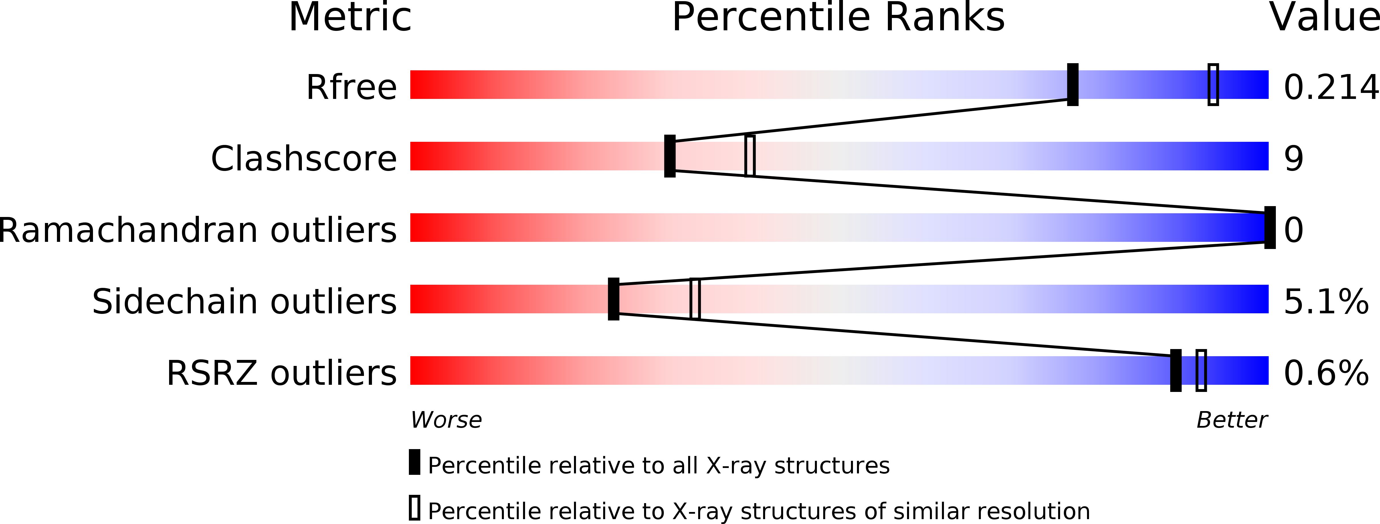
wwPDB Validation 3D Report Full Report
Entity ID: 1 | |||||
|---|---|---|---|---|---|
| Molecule | Chains | Sequence Length | Organism | Details | Image |
| Peptide ABC transporter, periplasmic peptide-binding protein | 529 | Vibrio cholerae O1 biovar El Tor str. N16961 | Mutation(s): 0 Gene Names: VC0620, VC_0620 | 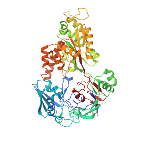 | |
UniProt | |||||
Find proteins for Q9KUA3 (Vibrio cholerae serotype O1 (strain ATCC 39315 / El Tor Inaba N16961)) Explore Q9KUA3 Go to UniProtKB: Q9KUA3 | |||||
Entity Groups | |||||
| Sequence Clusters | 30% Identity50% Identity70% Identity90% Identity95% Identity100% Identity | ||||
| UniProt Group | Q9KUA3 | ||||
Sequence AnnotationsExpand | |||||
| |||||
| Ligands 1 Unique | |||||
|---|---|---|---|---|---|
| ID | Chains | Name / Formula / InChI Key | 2D Diagram | 3D Interactions | |
| SO4 Query on SO4 | B [auth A], C [auth A], D [auth A] | SULFATE ION O4 S QAOWNCQODCNURD-UHFFFAOYSA-L |  | ||
| Length ( Å ) | Angle ( ˚ ) |
|---|---|
| a = 92.791 | α = 90 |
| b = 92.791 | β = 90 |
| c = 136.848 | γ = 120 |
| Software Name | Purpose |
|---|---|
| CNS | refinement |
| DENZO | data reduction |
| SCALEPACK | data scaling |
| CNS | phasing |
RCSB PDB (citation) is hosted by
RCSB PDB is a member of the

