4GET

Crystal structure of biogenic amine binding protein from Rhodnius prolixus


Experimental Data Snapshot

  • Method: X-RAY DIFFRACTION
  • Resolution: 2.24 Å
  • R-Value Free: 0.254 
  • R-Value Work: 0.185 
  • R-Value Observed: 0.188 

wwPDB Validation   3D Report Full Report


This is version 1.2 of the entry. See complete history


Literature

Structure and ligand-binding properties of the biogenic amine-binding protein from the saliva of a blood-feeding insect vector of Trypanosoma cruzi.

Xu, X.Chang, B.W.Mans, B.J.Ribeiro, J.M.Andersen, J.F.

(2013) Acta Crystallogr D Biol Crystallogr 69: 105-113

  • DOI: https://doi.org/10.1107/S0907444912043326
  • Primary Citation of Related Structures:  
    4GE1, 4GET, 4HFO

  • PubMed Abstract: 

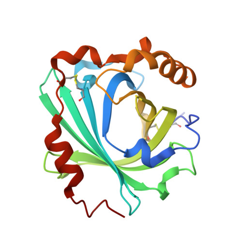
    Proteins that bind small-molecule mediators of inflammation and hemostasis are essential for blood-feeding by arthropod vectors of infectious disease. In ticks and triatomine insects, the lipocalin protein family is greatly expanded and members have been shown to bind biogenic amines, eicosanoids and ADP. These compounds are potent mediators of platelet activation, inflammation and vascular tone. In this paper, the structure of the amine-binding protein (ABP) from Rhodnius prolixus, a vector of the trypanosome that causes Chagas disease, is described. ABP binds the biogenic amines serotonin and norepinephrine with high affinity. A complex with tryptamine shows the presence of a binding site for a single ligand molecule in the central cavity of the β-barrel structure. The cavity contains significant additional volume, suggesting that this protein may have evolved from the related nitrophorin proteins, which bind a much larger heme ligand in the central cavity.


  • Organizational Affiliation

    Laboratory of Malaria and Vector Research, NIH/NIAID, Rockville, MD 20852, USA.


Macromolecules
Find similar proteins by:  (by identity cutoff)  |  3D Structure
Entity ID: 1
MoleculeChains Sequence LengthOrganismDetailsImage
Biogenic amine-binding protein
A, B, C, D
197Rhodnius prolixusMutation(s): 0 
UniProt
Find proteins for Q86PT9 (Rhodnius prolixus)
Explore Q86PT9 
Go to UniProtKB:  Q86PT9
Entity Groups  
Sequence Clusters30% Identity50% Identity70% Identity90% Identity95% Identity100% Identity
UniProt GroupQ86PT9
Sequence Annotations
Expand
  • Reference Sequence
Experimental Data & Validation

Experimental Data

  • Method: X-RAY DIFFRACTION
  • Resolution: 2.24 Å
  • R-Value Free: 0.254 
  • R-Value Work: 0.185 
  • R-Value Observed: 0.188 
  • Space Group: P 1 21 1
Unit Cell:
Length ( Å )Angle ( ˚ )
a = 68.727α = 90
b = 72.331β = 90.04
c = 79.491γ = 90
Software Package:
Software NamePurpose
DENZOdata reduction
SCALEPACKdata scaling
PHENIXrefinement
PDB_EXTRACTdata extraction
PHASERphasing

Structure Validation

View Full Validation Report


Entry History 

Deposition Data

Revision History  (Full details and data files)

  • Version 1.0: 2013-01-02
    Type: Initial release
  • Version 1.1: 2013-01-23
    Changes: Database references
  • Version 1.2: 2017-11-15
    Changes: Refinement description