4GBL

Crystal structure of aspart insulin at pH 8.5


Experimental Data Snapshot

  • Method: X-RAY DIFFRACTION
  • Resolution: 2.50 Å
  • R-Value Free: 0.262 
  • R-Value Work: 0.200 
  • R-Value Observed: 0.204 

wwPDB Validation   3D Report Full Report


This is version 1.3 of the entry. See complete history


Literature

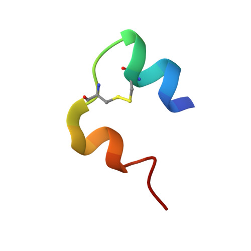
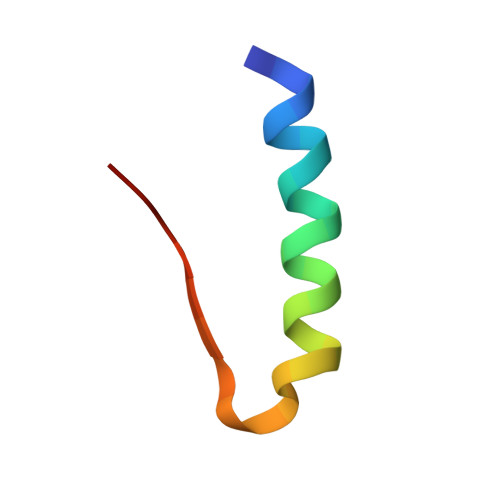
A T3R3 hexamer of the human insulin variant B28Asp.

Palmieri, L.C.Favero-Retto, M.P.Lourenco, D.Lima, L.M.

(2013) Biophys Chem 173: 1-7

  • DOI: https://doi.org/10.1016/j.bpc.2013.01.003
  • Primary Citation of Related Structures:  
    4GBC, 4GBI, 4GBK, 4GBL, 4GBN

  • PubMed Abstract: 

    Insulin shows a complex equilibrium between monomers and hexamers, involving varying conformers and association states. We sought to perform a structural characterization of the fast-acting human insulin variant B28Asp ("aspart"). Small-angle X-ray scattering measurements reveal similar globular behavior in both the aspart and regular human insulin, with a Rg of 19Å and a Dmax of approximately 50Å, indicating similar mean quaternary assembly distribution. Crystallographic assays revealed a T3R3 assembly of the aspart insulin formed by the TR dimer in the asymmetric unit, with all the first 8 residues of the B chain in the R-state monomer in helical conformation and the participation of its B3Asn in the stabilization of the hexamer. Our data provide access to novel structural information on aspart insulin such as an aspart insulin dimer in solution, the aspart insulin in T conformation and a pure R-state conformer establishing a T3R3 assembly, providing further insight on the stepwise conformational transition and assembly of this fast-insulin.


  • Organizational Affiliation

    School of Pharmacy, Federal University of Rio de Janeiro - UFRJ. CCS, Bss34, Ilha do Fundão, 21941-590, Rio de Janeiro, RJ, Brazil.


Macromolecules

Find similar proteins by:  Sequence   |   3D Structure  

Entity ID: 1
MoleculeChains Sequence LengthOrganismDetailsImage
Insulin A chain
A, C
21Homo sapiensMutation(s): 0 
UniProt & NIH Common Fund Data Resources
Find proteins for P01308 (Homo sapiens)
Explore P01308 
Go to UniProtKB:  P01308
PHAROS:  P01308
GTEx:  ENSG00000254647 
Entity Groups  
Sequence Clusters30% Identity50% Identity70% Identity90% Identity95% Identity100% Identity
UniProt GroupP01308
Sequence Annotations
Expand
  • Reference Sequence
Find similar proteins by:  (by identity cutoff)  |  3D Structure
Entity ID: 2
MoleculeChains Sequence LengthOrganismDetailsImage
Insulin B chain
B, D
30Homo sapiensMutation(s): 1 
UniProt & NIH Common Fund Data Resources
Find proteins for P01308 (Homo sapiens)
Explore P01308 
Go to UniProtKB:  P01308
PHAROS:  P01308
GTEx:  ENSG00000254647 
Entity Groups  
Sequence Clusters30% Identity50% Identity70% Identity90% Identity95% Identity100% Identity
UniProt GroupP01308
Sequence Annotations
Expand
  • Reference Sequence
Experimental Data & Validation

Experimental Data

  • Method: X-RAY DIFFRACTION
  • Resolution: 2.50 Å
  • R-Value Free: 0.262 
  • R-Value Work: 0.200 
  • R-Value Observed: 0.204 
  • Space Group: H 3
Unit Cell:
Length ( Å )Angle ( ˚ )
a = 78.57α = 90
b = 78.57β = 90
c = 37.73γ = 120
Software Package:
Software NamePurpose
MOSFLMdata reduction
SCALAdata scaling
MOLREPphasing
REFMACrefinement
PDB_EXTRACTdata extraction
Blu-Icedata collection
MAR345data collection

Structure Validation

View Full Validation Report


Entry History 

Deposition Data

Revision History  (Full details and data files)

  • Version 1.0: 2013-06-12
    Type: Initial release
  • Version 1.1: 2017-11-15
    Changes: Refinement description
  • Version 1.2: 2018-08-22
    Changes: Data collection, Database references, Derived calculations, Structure summary
  • Version 1.3: 2023-09-13
    Changes: Data collection, Database references, Derived calculations, Refinement description