4F9Z

Crystal Structure of human ERp27


Experimental Data Snapshot

  • Method: X-RAY DIFFRACTION
  • Resolution: 2.20 Å
  • R-Value Free: 0.217 
  • R-Value Work: 0.170 
  • R-Value Observed: 0.171 

wwPDB Validation   3D Report Full Report


This is version 1.2 of the entry. See complete history


Literature

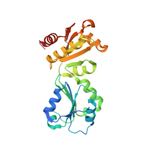
The Crystal Structure of the Protein-Disulfide Isomerase Family Member ERp27 Provides Insights into Its Substrate Binding Capabilities.

Kober, F.X.Koelmel, W.Kuper, J.Drechsler, J.Mais, C.Hermanns, H.M.Schindelin, H.

(2013) J Biol Chem 288: 2029-2039

  • DOI: https://doi.org/10.1074/jbc.M112.410522
  • Primary Citation of Related Structures:  
    4F9Z

  • PubMed Abstract: 

    About one-third of all cellular proteins pass through the secretory pathway and hence undergo oxidative folding in the endoplasmic reticulum (ER). Protein-disulfide isomerase (PDI) and related members of the PDI family assist in the folding of substrates by catalyzing the oxidation of two cysteines and isomerization of disulfide bonds as well as by acting as chaperones. In this study, we present the crystal structure of ERp27, a redox-inactive member of the PDI family. The structure reveals its substrate-binding cleft, which is homologous to PDI, but is able to adapt in size and hydrophobicity. Isothermal titration calorimetry experiments demonstrate that ERp27 is able to distinguish between folded and unfolded substrates, only interacting with the latter. ERp27 is up-regulated during ER stress, thus presumably allowing it to bind accumulating misfolded substrates and present them to ERp57 for catalysis.


  • Organizational Affiliation

    Rudolf Virchow Center for Experimental Biomedicine, University of Würzburg, Josef-Schneider-Strasse 2, 97080 Würzburg, Germany.


Macromolecules
Find similar proteins by:  (by identity cutoff)  |  3D Structure
Entity ID: 1
MoleculeChains Sequence LengthOrganismDetailsImage
Endoplasmic reticulum resident protein 27A [auth D],
B [auth A],
C [auth B],
D [auth C],
E
227Homo sapiensMutation(s): 0 
Gene Names: C12orf46ERP27UNQ781/PRO1575
UniProt & NIH Common Fund Data Resources
Find proteins for Q96DN0 (Homo sapiens)
Explore Q96DN0 
Go to UniProtKB:  Q96DN0
PHAROS:  Q96DN0
GTEx:  ENSG00000139055 
Entity Groups  
Sequence Clusters30% Identity50% Identity70% Identity90% Identity95% Identity100% Identity
UniProt GroupQ96DN0
Sequence Annotations
Expand
  • Reference Sequence
Small Molecules
Ligands 3 Unique
IDChains Name / Formula / InChI Key2D Diagram3D Interactions
PE3
Query on PE3

Download Ideal Coordinates CCD File 
M [auth B]3,6,9,12,15,18,21,24,27,30,33,36,39-TRIDECAOXAHENTETRACONTANE-1,41-DIOL
C28 H58 O15
ILLKMACMBHTSHP-UHFFFAOYSA-N
PE4
Query on PE4

Download Ideal Coordinates CCD File 
P [auth C]2-{2-[2-(2-{2-[2-(2-ETHOXY-ETHOXY)-ETHOXY]-ETHOXY}-ETHOXY)-ETHOXY]-ETHOXY}-ETHANOL
C16 H34 O8
PJWQOENWHPEPKI-UHFFFAOYSA-N
ACT
Query on ACT

Download Ideal Coordinates CCD File 
F [auth D]
G [auth D]
H [auth D]
I [auth D]
J [auth A]
F [auth D],
G [auth D],
H [auth D],
I [auth D],
J [auth A],
K [auth A],
L [auth A],
N [auth B],
O [auth B],
Q [auth C],
R [auth C],
S [auth E],
T [auth E]
ACETATE ION
C2 H3 O2
QTBSBXVTEAMEQO-UHFFFAOYSA-M
Experimental Data & Validation

Experimental Data

  • Method: X-RAY DIFFRACTION
  • Resolution: 2.20 Å
  • R-Value Free: 0.217 
  • R-Value Work: 0.170 
  • R-Value Observed: 0.171 
  • Space Group: P 1
Unit Cell:
Length ( Å )Angle ( ˚ )
a = 57.832α = 70.45
b = 68.359β = 88.16
c = 86.775γ = 64.96
Software Package:
Software NamePurpose
MxCuBEdata collection
SHELXSphasing
PHENIXrefinement
MOSFLMdata reduction
SCALAdata scaling

Structure Validation

View Full Validation Report


Entry History 

Deposition Data

Revision History  (Full details and data files)

  • Version 1.0: 2012-12-05
    Type: Initial release
  • Version 1.1: 2013-02-06
    Changes: Database references
  • Version 1.2: 2024-02-28
    Changes: Data collection, Database references, Derived calculations