4F88

X-ray Crystal Structure of PlyC


Experimental Data Snapshot

  • Method: X-RAY DIFFRACTION
  • Resolution: 3.30 Å
  • R-Value Free: 0.297 
  • R-Value Work: 0.266 
  • R-Value Observed: 0.267 

wwPDB Validation   3D Report Full Report


This is version 1.3 of the entry. See complete history


Literature

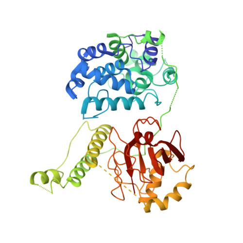
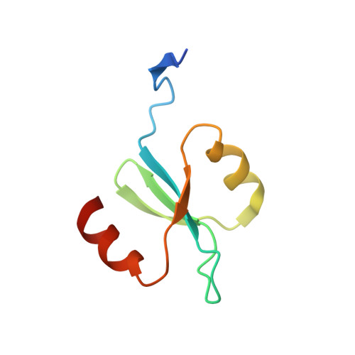
X-ray crystal structure of the streptococcal specific phage lysin PlyC.

McGowan, S.Buckle, A.M.Mitchell, M.S.Hoopes, J.T.Gallagher, D.T.Heselpoth, R.D.Shen, Y.Reboul, C.F.Law, R.H.Fischetti, V.A.Whisstock, J.C.Nelson, D.C.

(2012) Proc Natl Acad Sci U S A 109: 12752-12757

  • DOI: https://doi.org/10.1073/pnas.1208424109
  • Primary Citation of Related Structures:  
    4F87, 4F88

  • PubMed Abstract: 

    Bacteriophages deploy lysins that degrade the bacterial cell wall and facilitate virus egress from the host. When applied exogenously, these enzymes destroy susceptible microbes and, accordingly, have potential as therapeutic agents. The most potent lysin identified to date is PlyC, an enzyme assembled from two components (PlyCA and PlyCB) that is specific for streptococcal species. Here the structure of the PlyC holoenzyme reveals that a single PlyCA moiety is tethered to a ring-shaped assembly of eight PlyCB molecules. Structure-guided mutagenesis reveals that the bacterial cell wall binding is achieved through a cleft on PlyCB. Unexpectedly, our structural data reveal that PlyCA contains a glycoside hydrolase domain in addition to the previously recognized cysteine, histidine-dependent amidohydrolases/peptidases catalytic domain. The presence of eight cell wall-binding domains together with two catalytic domains may explain the extraordinary potency of the PlyC holoenyzme toward target bacteria.


  • Organizational Affiliation

    Department of Biochemistry and Molecular Biology, Monash University, Clayton 3800, Australia.


Macromolecules
Find similar proteins by:  (by identity cutoff)  |  3D Structure
Entity ID: 1
MoleculeChains Sequence LengthOrganismDetailsImage
PlyCAA [auth 1],
B [auth 2]
465Fischettivirus C1Mutation(s): 0 
Gene Names: orf11
UniProt
Find proteins for Q7Y3F1 (Streptococcus phage C1)
Explore Q7Y3F1 
Go to UniProtKB:  Q7Y3F1
Entity Groups  
Sequence Clusters30% Identity50% Identity70% Identity90% Identity95% Identity100% Identity
UniProt GroupQ7Y3F1
Sequence Annotations
Expand
  • Reference Sequence
Find similar proteins by:  (by identity cutoff)  |  3D Structure
Entity ID: 2
MoleculeChains Sequence LengthOrganismDetailsImage
PlyCB72Fischettivirus C1Mutation(s): 0 
Gene Names: orf9
UniProt
Find proteins for Q7Y3F3 (Streptococcus phage C1)
Explore Q7Y3F3 
Go to UniProtKB:  Q7Y3F3
Entity Groups  
Sequence Clusters30% Identity50% Identity70% Identity90% Identity95% Identity100% Identity
UniProt GroupQ7Y3F3
Sequence Annotations
Expand
  • Reference Sequence
Experimental Data & Validation

Experimental Data

  • Method: X-RAY DIFFRACTION
  • Resolution: 3.30 Å
  • R-Value Free: 0.297 
  • R-Value Work: 0.266 
  • R-Value Observed: 0.267 
  • Space Group: P 21 21 21
Unit Cell:
Length ( Å )Angle ( ˚ )
a = 93.853α = 90
b = 117.423β = 90
c = 222.192γ = 90
Software Package:
Software NamePurpose
HKL-2000data collection
PHASERphasing
BUSTERrefinement
HKL-2000data reduction
HKL-2000data scaling

Structure Validation

View Full Validation Report


Entry History 

Deposition Data

Revision History  (Full details and data files)

  • Version 1.0: 2012-07-25
    Type: Initial release
  • Version 1.1: 2012-08-01
    Changes: Database references
  • Version 1.2: 2012-08-15
    Changes: Database references
  • Version 1.3: 2023-09-13
    Changes: Data collection, Database references, Refinement description