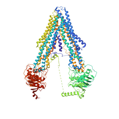
4F4C

The Crystal Structure of the Multi-Drug Transporter


Experimental Data Snapshot

  • Method: X-RAY DIFFRACTION
  • Resolution: 3.40 Å
  • R-Value Free: 0.283 
  • R-Value Work: 0.250 
  • R-Value Observed: 0.250 

wwPDB Validation   3D Report Full Report


Ligand Structure Quality Assessment 


This is version 2.1 of the entry. See complete history


Literature

Crystal structure of the multidrug transporter P-glycoprotein from Caenorhabditis elegans.

Jin, M.S.Oldham, M.L.Zhang, Q.Chen, J.

(2012) Nature 490: 566-569

  • DOI: https://doi.org/10.1038/nature11448
  • Primary Citation of Related Structures:  
    4F4C

  • PubMed Abstract: 

    P-glycoprotein (P-gp) is an ATP-binding cassette transporter that confers multidrug resistance in cancer cells. It also affects the absorption, distribution and clearance of cancer-unrelated drugs and xenobiotics. For these reasons, the structure and function of P-gp have been studied extensively for decades. Here we present biochemical characterization of P-gp from Caenorhabditis elegans and its crystal structure at a resolution of 3.4 ångströms. We find that the apparent affinities of P-gp for anticancer drugs actinomycin D and paclitaxel are approximately 4,000 and 100 times higher, respectively, in the membrane bilayer than in detergent. This affinity enhancement highlights the importance of membrane partitioning when a drug accesses the transporter in the membrane. Furthermore, the transporter in the crystal structure opens its drug pathway at the level of the membrane's inner leaflet. In the helices flanking the opening to the membrane, we observe extended loops that may mediate drug binding, function as hinges to gate the pathway or both. We also find that the interface between the transmembrane and nucleotide-binding domains, which couples ATP hydrolysis to transport, contains a ball-and-socket joint and salt bridges similar to the ATP-binding cassette importers, suggesting that ATP-binding cassette exporters and importers may use similar mechanisms to achieve alternating access for transport. Finally, a model of human P-gp derived from the structure of C. elegans P-gp not only is compatible with decades of biochemical analysis, but also helps to explain perplexing functional data regarding the Phe335Ala mutant. These results increase our understanding of the structure and function of this important molecule.


  • Organizational Affiliation

    Department of Biological Sciences, Purdue University, Indiana 47907, USA.


Macromolecules
Find similar proteins by:  (by identity cutoff)  |  3D Structure
Entity ID: 1
MoleculeChains Sequence LengthOrganismDetailsImage
Multidrug resistance protein pgp-11,321Caenorhabditis elegansMutation(s): 0 
Gene Names: pgp-1K08E7.9
EC: 3.6.3.44
Membrane Entity: Yes 
UniProt
Find proteins for P34712 (Caenorhabditis elegans)
Explore P34712 
Go to UniProtKB:  P34712
Entity Groups  
Sequence Clusters30% Identity50% Identity70% Identity90% Identity95% Identity100% Identity
UniProt GroupP34712
Sequence Annotations
Expand
  • Reference Sequence
Oligosaccharides

Help

Entity ID: 2
MoleculeChains Length2D Diagram Glycosylation3D Interactions
alpha-D-mannopyranose-(1-3)-beta-D-mannopyranose-(1-4)-2-acetamido-2-deoxy-beta-D-glucopyranose-(1-4)-2-acetamido-2-deoxy-beta-D-glucopyranose
B
4N-Glycosylation
Glycosylation Resources
GlyTouCan:  G81315DD
GlyCosmos:  G81315DD
GlyGen:  G81315DD
Small Molecules
Ligands 1 Unique
IDChains Name / Formula / InChI Key2D Diagram3D Interactions
0SA
Query on 0SA

Download Ideal Coordinates CCD File 
C [auth A],
D [auth A]
undecyl 4-O-alpha-D-glucopyranosyl-1-thio-beta-D-glucopyranoside
C23 H44 O10 S
SQISXDUZDUDUNY-GNKAUAAYSA-N
Experimental Data & Validation

Experimental Data

  • Method: X-RAY DIFFRACTION
  • Resolution: 3.40 Å
  • R-Value Free: 0.283 
  • R-Value Work: 0.250 
  • R-Value Observed: 0.250 
  • Space Group: P 21 21 21
Unit Cell:
Length ( Å )Angle ( ˚ )
a = 96.896α = 90
b = 155.36β = 90
c = 162.416γ = 90
Software Package:
Software NamePurpose
HKL-2000data collection
PHASERphasing
CNSrefinement
HKL-2000data reduction
HKL-2000data scaling

Structure Validation

View Full Validation Report


Ligand Structure Quality Assessment 


Entry History 

Deposition Data

Revision History  (Full details and data files)

  • Version 1.0: 2012-09-26
    Type: Initial release
  • Version 1.1: 2012-11-07
    Changes: Database references
  • Version 2.0: 2020-07-29
    Type: Remediation
    Reason: Carbohydrate remediation
    Changes: Advisory, Atomic model, Data collection, Derived calculations, Non-polymer description, Structure summary
  • Version 2.1: 2023-09-13
    Changes: Data collection, Database references, Refinement description, Structure summary