4F1Q

Human Artd8 (Parp14, Bal2) - catalytic domain in complex with A16(E)


Experimental Data Snapshot

  • Method: X-RAY DIFFRACTION
  • Resolution: 2.80 Å
  • R-Value Free: 0.283 
  • R-Value Work: 0.217 
  • R-Value Observed: 0.224 

wwPDB Validation   3D Report Full Report


Ligand Structure Quality Assessment 


This is version 1.2 of the entry. See complete history


Literature

Discovery of Ligands for ADP-Ribosyltransferases via Docking-Based Virtual Screening.

Andersson, C.D.Karlberg, T.Ekblad, T.Lindgren, A.E.Thorsell, A.G.Spjut, S.Uciechowska, U.Niemiec, M.S.Wittung-Stafshede, P.Weigelt, J.Elofsson, M.Schuler, H.Linusson, A.

(2012) J Med Chem 55: 7706-7718

  • DOI: https://doi.org/10.1021/jm300746d
  • Primary Citation of Related Structures:  
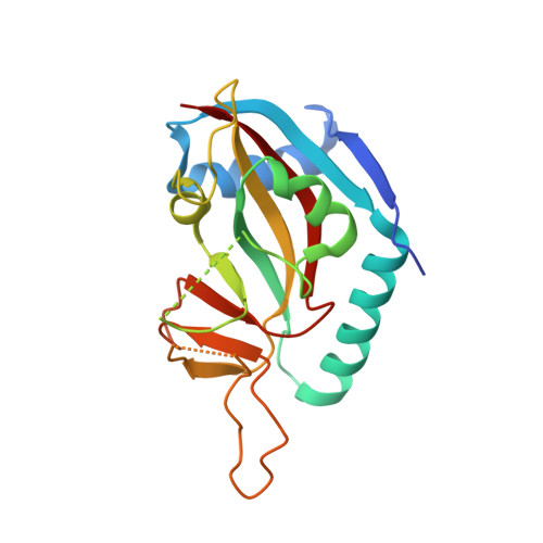
    4F0E, 4F1L, 4F1Q

  • PubMed Abstract: 

    The diphtheria toxin-like ADP-ribosyltransferases (ARTDs) are an enzyme family that catalyzes the transfer of ADP-ribose units onto substrate proteins by using nicotinamide adenine dinucleotide (NAD(+)) as a cosubstrate. They have a documented role in chromatin remodelling and DNA repair, and inhibitors of ARTD1 and 2 (PARP1 and 2) are currently in clinical trials for the treatment of cancer. The detailed function of most other ARTDs is still unknown. By using virtual screening, we identified small ligands of ARTD7 (PARP15/BAL3) and ARTD8 (PARP14/BAL2). Thermal-shift assays confirmed that 16 compounds, belonging to eight structural classes, bound to ARTD7/ARTD8. Affinity measurements with isothermal titration calorimetry for two isomers of the most promising hit compound confirmed binding in the low micromolar range to ARTD8. Crystal structures showed anchoring of the hits in the nicotinamide pocket. These results form a starting point in the development of chemical tools for the study of the role and function of ARTD7 and ARTD8.


  • Organizational Affiliation

    Department of Chemistry, Umeå University, SE-90187 Umeå, Sweden.


Macromolecules
Find similar proteins by:  (by identity cutoff)  |  3D Structure
Entity ID: 1
MoleculeChains Sequence LengthOrganismDetailsImage
Poly [ADP-ribose] polymerase 14
A, B
193Homo sapiensMutation(s): 0 
Gene Names: BAL2KIAA1268PARP14
EC: 2.4.2.30
UniProt & NIH Common Fund Data Resources
Find proteins for Q460N5 (Homo sapiens)
Explore Q460N5 
Go to UniProtKB:  Q460N5
PHAROS:  Q460N5
GTEx:  ENSG00000173193 
Entity Groups  
Sequence Clusters30% Identity50% Identity70% Identity90% Identity95% Identity100% Identity
UniProt GroupQ460N5
Sequence Annotations
Expand
  • Reference Sequence
Small Molecules
Binding Affinity Annotations 
IDSourceBinding Affinity
0RZ BindingDB:  4F1Q Kd: 1.12e+4 (nM) from 1 assay(s)
IC50: 2.00e+4 (nM) from 1 assay(s)
PDBBind:  4F1Q Kd: 1.12e+4 (nM) from 1 assay(s)
Experimental Data & Validation

Experimental Data

  • Method: X-RAY DIFFRACTION
  • Resolution: 2.80 Å
  • R-Value Free: 0.283 
  • R-Value Work: 0.217 
  • R-Value Observed: 0.224 
  • Space Group: C 2 2 21
Unit Cell:
Length ( Å )Angle ( ˚ )
a = 82.676α = 90
b = 144.986β = 90
c = 83.333γ = 90
Software Package:
Software NamePurpose
MxCuBEdata collection
MOLREPphasing
REFMACrefinement
XDSdata reduction
XSCALEdata scaling

Structure Validation

View Full Validation Report


Ligand Structure Quality Assessment 


Entry History 

Deposition Data

Revision History  (Full details and data files)

  • Version 1.0: 2012-09-05
    Type: Initial release
  • Version 1.1: 2012-09-26
    Changes: Database references
  • Version 1.2: 2023-09-13
    Changes: Data collection, Database references, Derived calculations, Refinement description