4EZN

Crystal structure of the substrate binding domain of E.coli DnaK in complex with pyrrhocoricin


Experimental Data Snapshot

  • Method: X-RAY DIFFRACTION
  • Resolution: 1.80 Å
  • R-Value Free: 0.238 
  • R-Value Work: 0.197 
  • R-Value Observed: 0.198 

wwPDB Validation   3D Report Full Report


This is version 1.3 of the entry. See complete history


Literature

Structural Studies on the Forward and Reverse Binding Modes of Peptides to the Chaperone DnaK.

Zahn, M.Berthold, N.Kieslich, B.Knappe, D.Hoffmann, R.Strater, N.

(2013) J Mol Biol 425: 2463-2479

  • DOI: https://doi.org/10.1016/j.jmb.2013.03.041
  • Primary Citation of Related Structures:  
    4EZN, 4EZO, 4EZP, 4EZQ, 4EZR, 4EZT, 4EZW, 4EZX, 4EZY, 4EZZ, 4F00, 4F01, 4HY9, 4HYB

  • PubMed Abstract: 

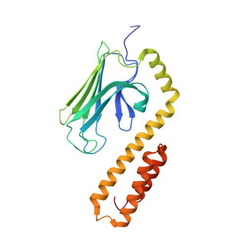
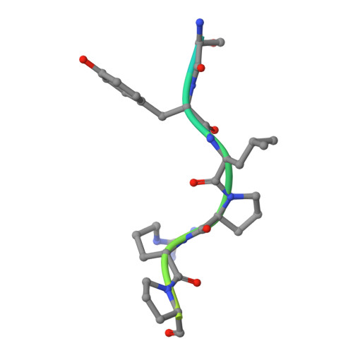
    Hsp70 chaperones have been implicated in assisting protein folding of newly synthesized polypeptide chains, refolding of misfolded proteins, and protein trafficking. For these functions, the chaperones need to exhibit a significant promiscuity in binding to different sequences of hydrophobic peptide stretches. To characterize the structural basis of sequence specificity and flexibility of the Escherichia coli Hsp70 chaperone DnaK, we have analyzed crystal structures of the substrate binding domain of the protein in complex with artificially designed peptides as well as small proline-rich antimicrobial peptides. The latter peptides from mammals and insects were identified to target DnaK after cell penetration. Interestingly, the complex crystal structures reveal two different peptide binding modes. The peptides can bind either in a forward or in a reverse direction to the conventional substrate binding cleft of DnaK in an extended conformation. Superposition of the two binding modes shows a remarkable similarity in the side chain orientations and hydrogen bonding pattern despite the reversed peptide orientation. The DnaK chaperone has evolved to bind peptides in both orientations in the substrate binding cleft with comparable energy without rearrangements of the protein. Optimal hydrophobic interactions with binding pockets -2 to 0 appear to be the main determinant for the orientation and sequence position of peptide binding.


  • Organizational Affiliation

    Institute of Bioanalytical Chemistry, Center for Biotechnology and Biomedicine, University of Leipzig, 04103 Leipzig, Germany.


Macromolecules
Find similar proteins by:  (by identity cutoff)  |  3D Structure
Entity ID: 1
MoleculeChains Sequence LengthOrganismDetailsImage
Chaperone protein DnaK
A, B
219Escherichia coli K-12Mutation(s): 0 
Gene Names: b0014dnaKgroPgrpFJW0013seg
UniProt
Find proteins for P0A6Y8 (Escherichia coli (strain K12))
Explore P0A6Y8 
Go to UniProtKB:  P0A6Y8
Entity Groups  
Sequence Clusters30% Identity50% Identity70% Identity90% Identity95% Identity100% Identity
UniProt GroupP0A6Y8
Sequence Annotations
Expand
  • Reference Sequence

Find similar proteins by:  Sequence   |   3D Structure  

Entity ID: 2
MoleculeChains Sequence LengthOrganismDetailsImage
Pyrrhocoricin
C, D
20Pyrrhocoris apterusMutation(s): 0 
UniProt
Find proteins for P37362 (Pyrrhocoris apterus)
Explore P37362 
Go to UniProtKB:  P37362
Entity Groups  
Sequence Clusters30% Identity50% Identity70% Identity90% Identity95% Identity100% Identity
UniProt GroupP37362
Sequence Annotations
Expand
  • Reference Sequence
Experimental Data & Validation

Experimental Data

  • Method: X-RAY DIFFRACTION
  • Resolution: 1.80 Å
  • R-Value Free: 0.238 
  • R-Value Work: 0.197 
  • R-Value Observed: 0.198 
  • Space Group: P 21 21 2
Unit Cell:
Length ( Å )Angle ( ˚ )
a = 77.58α = 90
b = 160.272β = 90
c = 44.928γ = 90
Software Package:
Software NamePurpose
MAR345data collection
REFMACrefinement
XDSdata reduction
SCALAdata scaling
REFMACphasing

Structure Validation

View Full Validation Report


Entry History 

Deposition Data

Revision History  (Full details and data files)

  • Version 1.0: 2013-04-17
    Type: Initial release
  • Version 1.1: 2013-07-17
    Changes: Database references
  • Version 1.2: 2017-11-15
    Changes: Refinement description
  • Version 1.3: 2024-02-28
    Changes: Data collection, Database references