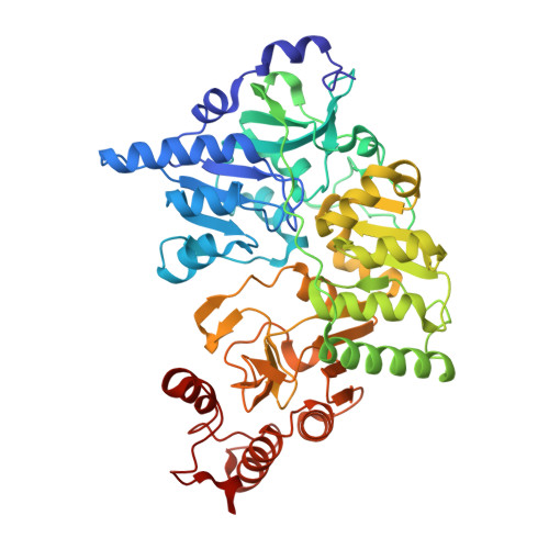
Crystal Structures of Acetobacter aceti Succinyl-Coenzyme A (CoA):Acetate CoA-Transferase Reveal Specificity Determinants and Illustrate the Mechanism Used by Class I CoA-Transferases.
Mullins, E.A., Kappock, T.J.(2012) Biochemistry 51: 8422-8434
- PubMed: 23030530
- DOI: https://doi.org/10.1021/bi300957f
- Primary Citation of Related Structures:
4EU3, 4EU4, 4EU5, 4EU6, 4EU7, 4EU8, 4EU9, 4EUA, 4EUB, 4EUC, 4EUD - PubMed Abstract:
Coenzyme A (CoA)-transferases catalyze transthioesterification reactions involving acyl-CoA substrates, using an active-site carboxylate to form covalent acyl anhydride and CoA thioester adducts. Mechanistic studies of class I CoA-transferases suggested that acyl-CoA binding energy is used to accelerate rate-limiting acyl transfers by compressing the substrate thioester tightly against the catalytic glutamate [White, H., and Jencks, W. P. (1976) J. Biol. Chem. 251, 1688-1699]. The class I CoA-transferase succinyl-CoA:acetate CoA-transferase is an acetic acid resistance factor (AarC) with a role in a variant citric acid cycle in Acetobacter aceti. In an effort to identify residues involved in substrate recognition, X-ray crystal structures of a C-terminally His(6)-tagged form (AarCH6) were determined for several wild-type and mutant complexes, including freeze-trapped acetylglutamyl anhydride and glutamyl-CoA thioester adducts. The latter shows the acetate product bound to an auxiliary site that is required for efficient carboxylate substrate recognition. A mutant in which the catalytic glutamate was changed to an alanine crystallized in a closed complex containing dethiaacetyl-CoA, which adopts an unusual curled conformation. A model of the acetyl-CoA Michaelis complex demonstrates the compression anticipated four decades ago by Jencks and reveals that the nucleophilic glutamate is held at a near-ideal angle for attack as the thioester oxygen is forced into an oxyanion hole composed of Gly388 NH and CoA N2″. CoA is nearly immobile along its entire length during all stages of the enzyme reaction. Spatial and sequence conservation of key residues indicates that this mechanism is general among class I CoA-transferases.
Organizational Affiliation:
Department of Biochemistry, Purdue University, West Lafayette, IN 47907-2063, USA.