


Crystal structure of ferric binding protein A
Wang, Q., Liu, X.Q., Wang, X.Q.To be published.
Experimental Data Snapshot
wwPDB Validation 3D Report Full Report

Entity ID: 1 | |||||
|---|---|---|---|---|---|
| Molecule | Chains | Sequence Length | Organism | Details | Image |
| Iron ABC transporter, periplasmic iron-binding protein | 336 | Thermus thermophilus HB8 | Mutation(s): 0 Gene Names: TTHA1628 | 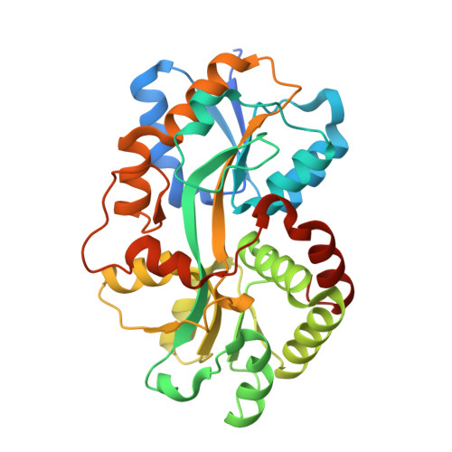 | |
UniProt | |||||
Find proteins for Q5SHV2 (Thermus thermophilus (strain ATCC 27634 / DSM 579 / HB8)) Explore Q5SHV2 Go to UniProtKB: Q5SHV2 | |||||
Entity Groups | |||||
| Sequence Clusters | 30% Identity50% Identity70% Identity90% Identity95% Identity100% Identity | ||||
| UniProt Group | Q5SHV2 | ||||
Sequence AnnotationsExpand | |||||
| |||||
| Ligands 2 Unique | |||||
|---|---|---|---|---|---|
| ID | Chains | Name / Formula / InChI Key | 2D Diagram | 3D Interactions | |
| CO3 Query on CO3 | D [auth A], F [auth B] | CARBONATE ION C O3 BVKZGUZCCUSVTD-UHFFFAOYSA-L |  | ||
| FE Query on FE | C [auth A], E [auth B] | FE (III) ION Fe VTLYFUHAOXGGBS-UHFFFAOYSA-N |  | ||
| Length ( Å ) | Angle ( ˚ ) |
|---|---|
| a = 63.602 | α = 90 |
| b = 63.602 | β = 90 |
| c = 266.701 | γ = 120 |
| Software Name | Purpose |
|---|---|
| HKL-2000 | data collection |
| PHASER | phasing |
| PHENIX | refinement |
| HKL-2000 | data reduction |
| HKL-2000 | data scaling |
RCSB PDB (citation) is hosted by
RCSB PDB is a member of the

