Mechanistic and structural analyses of the role of his67 in the yeast polyamine oxidase fms1.
Adachi, M.S., Taylor, A.B., Hart, P.J., Fitzpatrick, P.F.(2012) Biochemistry 51: 4888-4897
- PubMed: 22642831
- DOI: https://doi.org/10.1021/bi300517s
- Primary Citation of Related Structures:
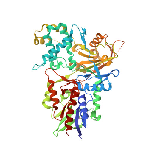
4ECH - PubMed Abstract:
The flavoprotein oxidase Fms1 from Saccharomyces cerevisiae catalyzes the oxidation of spermine and N(1)-acetylspermine to spermidine and 3-aminopropanal or N-acetyl-3-aminopropanal. Within the active site of Fms1, His67 is positioned to form hydrogen bonds with the polyamine substrate. This residue is also conserved in other polyamine oxidases. The catalytic properties of H67Q, H67N, and H67A Fms1 have been characterized to evaluate the role of this residue in catalysis. With both spermine and N(1)-acetylspermine as the amine substrate, the value of the first-order rate constant for flavin reduction decreases 2-3 orders of magnitude, with the H67Q mutation having the smallest effect and H67N the largest. The k(cat)/K(O2) value changes very little upon mutation with N(1)-acetylspermine as the amine substrate and decreases only an order of magnitude with spermine. The k(cat)/K(M)-pH profiles with N(1)-acetylspermine are bell-shaped for all the mutants; the similarity to the profile of the wild-type enzyme rules out His67 as being responsible for either of the pK(a) values. The pH profiles for the rate constant for flavin reduction for all the mutant enzymes similarly show the same pK(a) as wild-type Fms1, about ∼7.4; this pK(a) is assigned to the substrate N4. The k(cat)/K(O2)-pH profiles for wild-type Fms1 and the H67A enzyme both show a pK(a) of about ∼6.9; this suggests His67 is not responsible for this pH behavior. With the H67Q, H67N, and H67A enzymes the k(cat) value decreases when a single residue is protonated, as is the case with the wild-type enzyme. The structure of H67Q Fms1 has been determined at a resolution of 2.4 Å. The structure shows that the mutation disrupts a hydrogen bond network in the active site, suggesting that His67 is important both for direct interactions with the substrate and to maintain the overall active site structure.
Organizational Affiliation:
Department of Biochemistry, University of Texas Health Science Center, San Antonio, Texas 78229, USA.