4E0V

Structure of L-amino acid oxidase from the B. jararacussu venom


Experimental Data Snapshot

  • Method: X-RAY DIFFRACTION
  • Resolution: 3.10 Å
  • R-Value Free: 0.259 
  • R-Value Work: 0.181 
  • R-Value Observed: 0.185 

wwPDB Validation   3D Report Full Report


Ligand Structure Quality Assessment 


This is version 1.1 of the entry. See complete history


Literature

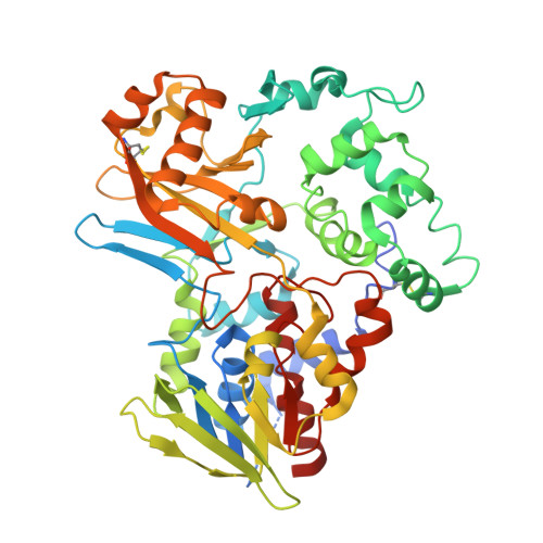
Structural insights into selectivity and cofactor binding in snake venom L-amino acid oxidases.

Ullah, A.Souza, T.A.Abrego, J.R.Betzel, C.Murakami, M.T.Arni, R.K.

(2012) Biochem Biophys Res Commun 421: 124-128

  • DOI: https://doi.org/10.1016/j.bbrc.2012.03.129
  • Primary Citation of Related Structures:  
    4E0V

  • PubMed Abstract: 

    L-Amino acid oxidases (LAAOs) are flavoenzymes that catalytically deaminate L-amino acids to corresponding α-keto acids with the concomitant production of ammonia (NH(3)) and hydrogen peroxide (H(2)O(2)). Particularly, snake venom LAAOs have been attracted much attention due to their diverse clinical and biological effects, interfering on human coagulation factors and being cytotoxic against some pathogenic bacteria and Leishmania ssp. In this work, a new LAAO from Bothrops jararacussu venom (BjsuLAAO) was purified, functionally characterized and its structure determined by X-ray crystallography at 3.1 Å resolution. BjsuLAAO showed high catalytic specificity for aromatic and aliphatic large side-chain amino acids. Comparative structural analysis with prokaryotic LAAOs, which exhibit low specificity, indicates the importance of the active-site volume in modulating enzyme selectivity. Surprisingly, the flavin adenine dinucleotide (FAD) cofactor was found in a different orientation canonically described for both prokaryotic and eukaryotic LAAOs. In this new conformational state, the adenosyl group is flipped towards the 62-71 loop, being stabilized by several hydrogen-bond interactions, which is equally stable to the classical binding mode.


  • Organizational Affiliation

    Centro Multiusuário de Inovação Biomolecular, Departamento de Física, Universidade Estadual Paulista (UNESP), 15054-000 São José do Rio Preto, SP, Brazil.


Macromolecules
Find similar proteins by:  (by identity cutoff)  |  3D Structure
Entity ID: 1
MoleculeChains Sequence LengthOrganismDetailsImage
L-amino-acid oxidase
A, B
497Bothrops jararacussuMutation(s): 0 
EC: 1.4.3.2
UniProt
Find proteins for Q6TGQ9 (Bothrops jararacussu)
Explore Q6TGQ9 
Go to UniProtKB:  Q6TGQ9
Entity Groups  
Sequence Clusters30% Identity50% Identity70% Identity90% Identity95% Identity100% Identity
UniProt GroupQ6TGQ9
Sequence Annotations
Expand
  • Reference Sequence
Small Molecules
Ligands 1 Unique
IDChains Name / Formula / InChI Key2D Diagram3D Interactions
FAD
Query on FAD

Download Ideal Coordinates CCD File 
C [auth A],
D [auth B]
FLAVIN-ADENINE DINUCLEOTIDE
C27 H33 N9 O15 P2
VWWQXMAJTJZDQX-UYBVJOGSSA-N
Experimental Data & Validation

Experimental Data

  • Method: X-RAY DIFFRACTION
  • Resolution: 3.10 Å
  • R-Value Free: 0.259 
  • R-Value Work: 0.181 
  • R-Value Observed: 0.185 
  • Space Group: P 1 21 1
Unit Cell:
Length ( Å )Angle ( ˚ )
a = 66.385α = 90
b = 72.19β = 90.9
c = 101.527γ = 90
Software Package:
Software NamePurpose
MOLREPphasing
REFMACrefinement
HKL-2000data reduction
HKL-2000data scaling

Structure Validation

View Full Validation Report


Ligand Structure Quality Assessment 


Entry History 

Deposition Data

Revision History  (Full details and data files)

  • Version 1.0: 2012-04-25
    Type: Initial release
  • Version 1.1: 2012-08-15
    Changes: Database references