4E0S

Crystal Structure of C5b-6


Experimental Data Snapshot

  • Method: X-RAY DIFFRACTION
  • Resolution: 4.21 Å
  • R-Value Free: 0.278 
  • R-Value Work: 0.218 
  • R-Value Observed: 0.221 

wwPDB Validation   3D Report Full Report


Ligand Structure Quality Assessment 


This is version 2.1 of the entry. See complete history


Literature

Crystal structure of c5b-6 suggests structural basis for priming assembly of the membrane attack complex.

Aleshin, A.E.Discipio, R.G.Stec, B.Liddington, R.C.

(2012) J Biol Chem 287: 19642-19652

  • DOI: https://doi.org/10.1074/jbc.M112.361121
  • Primary Citation of Related Structures:  
    4E0S

  • PubMed Abstract: 

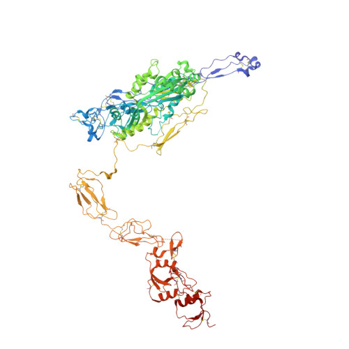
    The complement membrane attack complex (MAC) forms transmembrane pores in pathogen membranes. The first step in MAC assembly is cleavage of C5 to generate metastable C5b, which forms a stable complex with C6, termed C5b-6. C5b-6 initiates pore formation via the sequential recruitment of homologous proteins: C7, C8, and 12-18 copies of C9, each of which comprises a central MAC-perforin domain flanked by auxiliary domains. We recently proposed a model of pore assembly, in which the auxiliary domains play key roles, both in stabilizing the closed conformation of the protomers and in driving the sequential opening of the MAC-perforin β-sheet of each new recruit to the growing pore. Here, we describe an atomic model of C5b-6 at 4.2 Å resolution. We show that C5b provides four interfaces for the auxiliary domains of C6. The largest interface is created by the insertion of an interdomain linker from C6 into a hydrophobic groove created by a major reorganization of the α-helical domain of C5b. In combination with the rigid body docking of N-terminal elements of both proteins, C5b becomes locked into a stable conformation. Both C6 auxiliary domains flanking the linker pack tightly against C5b. The net effect is to induce the clockwise rigid body rotation of four auxiliary domains, as well as the opening/twisting of the central β-sheet of C6, in the directions predicted by our model to activate or prime C6 for the subsequent steps in MAC assembly. The complex also suggests novel small molecule strategies for modulating pathological MAC assembly.


  • Organizational Affiliation

    Program on Infectious Diseases, Sanford-Burnham Medical Research Institute, La Jolla, California 92037, USA.


Macromolecules
Find similar proteins by:  (by identity cutoff)  |  3D Structure
Entity ID: 1
MoleculeChains Sequence LengthOrganismDetailsImage
Complement C51,676Homo sapiensMutation(s): 0 
UniProt & NIH Common Fund Data Resources
Find proteins for P01031 (Homo sapiens)
Explore P01031 
Go to UniProtKB:  P01031
PHAROS:  P01031
GTEx:  ENSG00000106804 
Entity Groups  
Sequence Clusters30% Identity50% Identity70% Identity90% Identity95% Identity100% Identity
UniProt GroupP01031
Sequence Annotations
Expand
  • Reference Sequence
Find similar proteins by:  (by identity cutoff)  |  3D Structure
Entity ID: 2
MoleculeChains Sequence LengthOrganismDetailsImage
Complement component C6913Homo sapiensMutation(s): 0 
UniProt & NIH Common Fund Data Resources
Find proteins for P13671 (Homo sapiens)
Explore P13671 
Go to UniProtKB:  P13671
PHAROS:  P13671
GTEx:  ENSG00000039537 
Entity Groups  
Sequence Clusters30% Identity50% Identity70% Identity90% Identity95% Identity100% Identity
UniProt GroupP13671
Sequence Annotations
Expand
  • Reference Sequence
Oligosaccharides

Help

Entity ID: 3
MoleculeChains Length2D Diagram Glycosylation3D Interactions
2-acetamido-2-deoxy-beta-D-glucopyranose-(1-4)-2-acetamido-2-deoxy-beta-D-glucopyranose
C, D
2N-Glycosylation
Glycosylation Resources
GlyTouCan:  G42666HT
GlyCosmos:  G42666HT
GlyGen:  G42666HT
Entity ID: 4
MoleculeChains Length2D Diagram Glycosylation3D Interactions
beta-D-glucopyranose-(1-3)-alpha-L-fucopyranose
E
2O-Glycosylation
Glycosylation Resources
GlyTouCan:  G36855WW
GlyCosmos:  G36855WW
GlyGen:  G36855WW
Experimental Data & Validation

Experimental Data

  • Method: X-RAY DIFFRACTION
  • Resolution: 4.21 Å
  • R-Value Free: 0.278 
  • R-Value Work: 0.218 
  • R-Value Observed: 0.221 
  • Space Group: I 21 21 21
Unit Cell:
Length ( Å )Angle ( ˚ )
a = 158.949α = 90
b = 227.529β = 90
c = 278.157γ = 90
Software Package:
Software NamePurpose
Blu-Icedata collection
PHASERphasing
REFMACrefinement
HKL-2000data reduction
HKL-2000data scaling

Structure Validation

View Full Validation Report


Ligand Structure Quality Assessment 


Entry History 

Deposition Data

Revision History  (Full details and data files)

  • Version 1.0: 2012-04-18
    Type: Initial release
  • Version 1.1: 2012-04-25
    Changes: Database references
  • Version 1.2: 2012-06-20
    Changes: Database references
  • Version 1.3: 2017-11-15
    Changes: Refinement description
  • Version 2.0: 2020-07-29
    Type: Remediation
    Reason: Carbohydrate remediation
    Changes: Advisory, Atomic model, Data collection, Derived calculations, Structure summary
  • Version 2.1: 2023-09-13
    Changes: Data collection, Database references, Refinement description, Structure summary