


Crystal structure of MutT NUDIX hydrolase from Burkholderia pseudomallei
Edwards, T.E., Clifton, M.C., Seattle Structural Genomics Center for Infectious Disease (SSGCID)To be published.
Experimental Data Snapshot
wwPDB Validation 3D Report Full Report

Entity ID: 1 | |||||
|---|---|---|---|---|---|
| Molecule | Chains | Sequence Length | Organism | Details | Image |
| MutT/NUDIX family protein | 157 | Burkholderia pseudomallei 1710b | Mutation(s): 0 Gene Names: BURPS1710b_0547 | 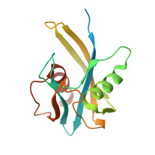 | |
UniProt | |||||
Find proteins for Q3JWU2 (Burkholderia pseudomallei (strain 1710b)) Explore Q3JWU2 Go to UniProtKB: Q3JWU2 | |||||
Entity Groups | |||||
| Sequence Clusters | 30% Identity50% Identity70% Identity90% Identity95% Identity100% Identity | ||||
| UniProt Group | Q3JWU2 | ||||
Sequence AnnotationsExpand | |||||
| |||||
| Length ( Å ) | Angle ( ˚ ) |
|---|---|
| a = 78.04 | α = 90 |
| b = 78.04 | β = 90 |
| c = 113.92 | γ = 90 |
| Software Name | Purpose |
|---|---|
| XSCALE | data scaling |
| PHASER | phasing |
| REFMAC | refinement |
| PDB_EXTRACT | data extraction |
| XDS | data reduction |
RCSB PDB (citation) is hosted by
RCSB PDB is a member of the

