4D51

Crystal structure of CymA from Klebsiella oxytoca


Experimental Data Snapshot

  • Method: X-RAY DIFFRACTION
  • Resolution: 2.30 Å
  • R-Value Free: 0.280 
  • R-Value Work: 0.214 
  • R-Value Observed: 0.217 

wwPDB Validation   3D Report Full Report


Ligand Structure Quality Assessment 


This is version 1.4 of the entry. See complete history


Literature

Outer-membrane translocation of bulky small molecules by passive diffusion.

van den Berg, B.Prathyusha Bhamidimarri, S.Dahyabhai Prajapati, J.Kleinekathofer, U.Winterhalter, M.

(2015) Proc Natl Acad Sci U S A 112: E2991-E2999

  • DOI: https://doi.org/10.1073/pnas.1424835112
  • Primary Citation of Related Structures:  
    4D51, 4D5B, 4D5D, 4V3G, 4V3H

  • PubMed Abstract: 

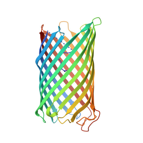
    The outer membrane (OM) of gram-negative bacteria forms a protective layer around the cell that serves as a permeability barrier to prevent unrestricted access of noxious substances. The permeability barrier of the OM results partly from the limited pore diameters of OM diffusion channels. As a consequence, there is an "OM size-exclusion limit," and the uptake of bulky molecules with molecular masses of more than ∼ 600 Da is thought to be mediated by TonB-dependent, active transporters. Intriguingly, the OM protein CymA from Klebsiella oxytoca does not depend on TonB but nevertheless mediates efficient OM passage of cyclodextrins with diameters of up to ∼ 15 Å. Here we show, by using X-ray crystallography, molecular dynamics simulations, and single-channel electrophysiology, that CymA forms a monomeric 14-stranded β-barrel with a large pore that is occluded on the periplasmic side by the N-terminal 15 residues of the protein. Representing a previously unidentified paradigm in OM transport, CymA mediates the passive diffusion of bulky molecules via an elegant transport mechanism in which a mobile element formed by the N terminus acts as a ligand-expelled gate to preserve the permeability barrier of the OM.


  • Organizational Affiliation

    Institute for Cell and Molecular Biosciences, The Medical School, Newcastle University, Newcastle upon Tyne NE2 4HH, United Kingdom; bert.van-den-berg@ncl.ac.uk.


Macromolecules
Find similar proteins by:  (by identity cutoff)  |  3D Structure
Entity ID: 1
MoleculeChains Sequence LengthOrganismDetailsImage
CYMA324Klebsiella oxytocaMutation(s): 0 
Membrane Entity: Yes 
UniProt
Find proteins for Q48391 (Klebsiella oxytoca)
Explore Q48391 
Go to UniProtKB:  Q48391
Entity Groups  
Sequence Clusters30% Identity50% Identity70% Identity90% Identity95% Identity100% Identity
UniProt GroupQ48391
Sequence Annotations
Expand
  • Reference Sequence
Small Molecules
Ligands 1 Unique
IDChains Name / Formula / InChI Key2D Diagram3D Interactions
C8E
Query on C8E

Download Ideal Coordinates CCD File 
B [auth A](HYDROXYETHYLOXY)TRI(ETHYLOXY)OCTANE
C16 H34 O5
FEOZZFHAVXYAMB-UHFFFAOYSA-N
Experimental Data & Validation

Experimental Data

  • Method: X-RAY DIFFRACTION
  • Resolution: 2.30 Å
  • R-Value Free: 0.280 
  • R-Value Work: 0.214 
  • R-Value Observed: 0.217 
  • Space Group: C 2 2 21
Unit Cell:
Length ( Å )Angle ( ˚ )
a = 59.62α = 90
b = 228.12β = 90
c = 60.61γ = 90
Software Package:
Software NamePurpose
PHENIXrefinement
XDSdata reduction
Aimlessdata scaling
PHASERphasing

Structure Validation

View Full Validation Report


Ligand Structure Quality Assessment 


Entry History 

Deposition Data

Revision History  (Full details and data files)

  • Version 1.0: 2015-06-03
    Type: Initial release
  • Version 1.1: 2015-06-10
    Changes: Database references, Source and taxonomy
  • Version 1.2: 2015-06-24
    Changes: Database references
  • Version 1.3: 2018-02-21
    Changes: Advisory, Database references, Structure summary
  • Version 1.4: 2023-12-20
    Changes: Advisory, Data collection, Database references, Derived calculations, Other, Refinement description