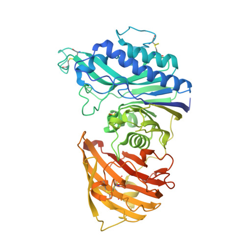
4CYF

The structure of vanin-1: defining the link between metabolic disease, oxidative stress and inflammation


Experimental Data Snapshot

  • Method: X-RAY DIFFRACTION
  • Resolution: 2.25 Å
  • R-Value Free: 0.222 
  • R-Value Work: 0.195 
  • R-Value Observed: 0.197 

wwPDB Validation   3D Report Full Report


Ligand Structure Quality Assessment 


This is version 3.0 of the entry. See complete history


Literature

The Structure of Vanin-1: A Key Enzyme Linking Metabolic Disease and Inflammation

Boersma, Y.L.Newman, J.Adams, T.E.Cowieson, N.Krippner, G.Bozaoglu, K.Peat, T.S.

(2014) Acta Crystallogr D Biol Crystallogr 70: 3320

  • DOI: https://doi.org/10.1107/S1399004714022767
  • Primary Citation of Related Structures:  
    4CYF, 4CYG, 4CYY

  • PubMed Abstract: 

    Although part of the coenzyme A pathway, vanin 1 (also known as pantetheinase) sits on the cell surface of many cell types as an ectoenzyme, catalyzing the breakdown of pantetheine to pantothenic acid (vitamin B5) and cysteamine, a strong reducing agent. Vanin 1 was initially discovered as a protein involved in the homing of leukocytes to the thymus. Numerous studies have shown that vanin 1 is involved in inflammation, and more recent studies have shown a key role in metabolic disease. Here, the X-ray crystal structure of human vanin 1 at 2.25 Å resolution is presented, which is the first reported structure from the vanin family, as well as a crystal structure of vanin 1 bound to a specific inhibitor. These structures illuminate how vanin 1 can mediate its biological roles by way of both enzymatic activity and protein-protein interactions. Furthermore, it sheds light on how the enzymatic activity is regulated by a novel allosteric mechanism at a domain interface.


  • Organizational Affiliation

    Department of Pharmaceutical Biology, Groningen Research Institute of Pharmacy, University of Groningen, A. Deusinglaan 1, 9713 AV Groningen, The Netherlands.


Macromolecules
Find similar proteins by:  (by identity cutoff)  |  3D Structure
Entity ID: 1
MoleculeChains Sequence LengthOrganismDetailsImage
PANTETHEINASE
A, B
506Homo sapiensMutation(s): 1 
EC: 3.5.1.92
UniProt & NIH Common Fund Data Resources
Find proteins for O95497 (Homo sapiens)
Explore O95497 
Go to UniProtKB:  O95497
PHAROS:  O95497
GTEx:  ENSG00000112299 
Entity Groups  
Sequence Clusters30% Identity50% Identity70% Identity90% Identity95% Identity100% Identity
UniProt GroupO95497
Sequence Annotations
Expand
  • Reference Sequence
Oligosaccharides

Help

Entity ID: 2
MoleculeChains Length2D Diagram Glycosylation3D Interactions
2-acetamido-2-deoxy-beta-D-glucopyranose-(1-4)-2-acetamido-2-deoxy-beta-D-glucopyranose
C
2N-Glycosylation
Glycosylation Resources
GlyTouCan:  G42666HT
GlyCosmos:  G42666HT
GlyGen:  G42666HT
Experimental Data & Validation

Experimental Data

  • Method: X-RAY DIFFRACTION
  • Resolution: 2.25 Å
  • R-Value Free: 0.222 
  • R-Value Work: 0.195 
  • R-Value Observed: 0.197 
  • Space Group: P 43 21 2
Unit Cell:
Length ( Å )Angle ( ˚ )
a = 119.941α = 90
b = 119.941β = 90
c = 221.887γ = 90
Software Package:
Software NamePurpose
REFMACrefinement
XDSdata reduction
SCALAdata scaling
Auto-Rickshawphasing

Structure Validation

View Full Validation Report


Ligand Structure Quality Assessment 


Entry History 

Deposition Data

Revision History  (Full details and data files)

  • Version 1.0: 2014-12-10
    Type: Initial release
  • Version 1.1: 2015-02-25
    Changes: Database references
  • Version 2.0: 2017-06-14
    Changes: Advisory, Atomic model, Derived calculations
  • Version 2.1: 2019-05-08
    Changes: Data collection, Experimental preparation
  • Version 3.0: 2020-07-29
    Type: Remediation
    Reason: Carbohydrate remediation
    Changes: Advisory, Atomic model, Data collection, Derived calculations, Other, Structure summary