4CXV

Structure of bifunctional endonuclease (AtBFN2) in complex with phosphate.


Experimental Data Snapshot

  • Method: X-RAY DIFFRACTION
  • Resolution: 2.00 Å
  • R-Value Free: 0.249 
  • R-Value Work: 0.202 
  • R-Value Observed: 0.204 

wwPDB Validation   3D Report Full Report


Ligand Structure Quality Assessment 


This is version 2.1 of the entry. See complete history


Literature

Structural Insights of the Ssdna Binding Site in the Multifunctional Endonuclease Atbfn2 from Arabidopsis Thaliana.

Yu, T.Maestre-Reyna, M.Ko, C.Ko, T.Sun, Y.Lin, T.Shaw, J.Wang, A.H.

(2014) PLoS One 9: 05821

  • DOI: https://doi.org/10.1371/journal.pone.0105821
  • Primary Citation of Related Structures:  
    4CWM, 4CXO, 4CXP, 4CXV

  • PubMed Abstract: 

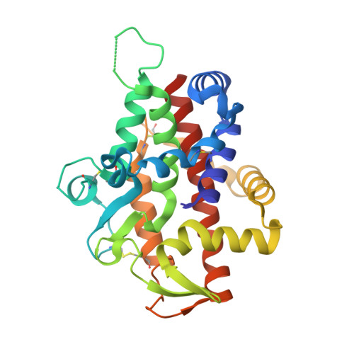
    The multi S1/P1 nuclease AtBFN2 (EC 3.1.30.1) encoded by the Arabidopsis thaliana At1g68290 gene is a glycoprotein that digests RNA, ssDNA, and dsDNA. AtBFN2 depends on three zinc ions for cleaving DNA and RNA at 3'-OH to yield 5'-nucleotides. In addition, AtBFN2's enzymatic activity is strongly glycan dependent. Plant Zn(2+)-dependent endonucleases present a unique fold, and belong to the Phospholipase C (PLC)/P1 nuclease superfamily. In this work, we present the first complete, ligand-free, AtBFN2 crystal structure, along with sulfate, phosphate and ssDNA co-crystal structures. With these, we were able to provide better insight into the glycan structure and possible enzymatic mechanism. In comparison with other nucleases, the AtBFN2/ligand-free and AtBFN2/PO4 models suggest a similar, previously proposed, catalytic mechanism. Our data also confirm that the phosphate and vanadate can inhibit the enzyme activity by occupying the active site. More importantly, the AtBFN2/A5T structure reveals a novel and conserved secondary binding site, which seems to be important for plant Zn(2+)-dependent endonucleases. Based on these findings, we propose a rational ssDNA binding model, in which the ssDNA wraps itself around the protein and the attached surface glycan, in turn, reinforces the binding complex.


  • Organizational Affiliation

    Institute of Bioinformatics and Structural Biology, National Tsing Hua University, Hsinchu, Taiwan; Institute of Biological Chemistry, Academia Sinica, Taipei, Taiwan.


Macromolecules
Find similar proteins by:  (by identity cutoff)  |  3D Structure
Entity ID: 1
MoleculeChains Sequence LengthOrganismDetailsImage
ENDONUCLEASE 2
A, B
269Arabidopsis thalianaMutation(s): 0 
EC: 3.1.30.1
UniProt
Find proteins for Q9C9G4 (Arabidopsis thaliana)
Explore Q9C9G4 
Go to UniProtKB:  Q9C9G4
Entity Groups  
Sequence Clusters30% Identity50% Identity70% Identity90% Identity95% Identity100% Identity
UniProt GroupQ9C9G4
Sequence Annotations
Expand
  • Reference Sequence
Oligosaccharides

Help

Entity ID: 2
MoleculeChains Length2D Diagram Glycosylation3D Interactions
alpha-D-mannopyranose-(1-3)-beta-D-mannopyranose-(1-4)-2-acetamido-2-deoxy-beta-D-glucopyranose-(1-4)-2-acetamido-2-deoxy-beta-D-glucopyranose
C
4N-Glycosylation
Glycosylation Resources
GlyTouCan:  G81315DD
GlyCosmos:  G81315DD
GlyGen:  G81315DD
Entity ID: 3
MoleculeChains Length2D Diagram Glycosylation3D Interactions
2-acetamido-2-deoxy-beta-D-glucopyranose-(1-4)-2-acetamido-2-deoxy-beta-D-glucopyranose
D, F
2N-Glycosylation
Glycosylation Resources
GlyTouCan:  G42666HT
GlyCosmos:  G42666HT
GlyGen:  G42666HT
Entity ID: 4
MoleculeChains Length2D Diagram Glycosylation3D Interactions
alpha-D-mannopyranose-(1-6)-alpha-D-mannopyranose-(1-6)-[alpha-D-mannopyranose-(1-3)]beta-D-mannopyranose-(1-4)-2-acetamido-2-deoxy-beta-D-glucopyranose-(1-4)-2-acetamido-2-deoxy-beta-D-glucopyranose
E
6N-Glycosylation
Glycosylation Resources
GlyTouCan:  G34442SS
GlyCosmos:  G34442SS
GlyGen:  G34442SS
Entity ID: 5
MoleculeChains Length2D Diagram Glycosylation3D Interactions
beta-D-mannopyranose-(1-4)-2-acetamido-2-deoxy-beta-D-glucopyranose-(1-4)-2-acetamido-2-deoxy-beta-D-glucopyranose
G
3N-Glycosylation
Glycosylation Resources
GlyTouCan:  G15407YE
GlyCosmos:  G15407YE
GlyGen:  G15407YE
Small Molecules
Ligands 3 Unique
IDChains Name / Formula / InChI Key2D Diagram3D Interactions
NAG
Query on NAG

Download Ideal Coordinates CCD File 
H [auth A]2-acetamido-2-deoxy-beta-D-glucopyranose
C8 H15 N O6
OVRNDRQMDRJTHS-FMDGEEDCSA-N
PO4
Query on PO4

Download Ideal Coordinates CCD File 
I [auth A],
M [auth B]
PHOSPHATE ION
O4 P
NBIIXXVUZAFLBC-UHFFFAOYSA-K
ZN
Query on ZN

Download Ideal Coordinates CCD File 
J [auth A]
K [auth A]
L [auth A]
N [auth B]
O [auth B]
J [auth A],
K [auth A],
L [auth A],
N [auth B],
O [auth B],
P [auth B]
ZINC ION
Zn
PTFCDOFLOPIGGS-UHFFFAOYSA-N
Experimental Data & Validation

Experimental Data

  • Method: X-RAY DIFFRACTION
  • Resolution: 2.00 Å
  • R-Value Free: 0.249 
  • R-Value Work: 0.202 
  • R-Value Observed: 0.204 
  • Space Group: P 1
Unit Cell:
Length ( Å )Angle ( ˚ )
a = 45.524α = 71.34
b = 52.87β = 78.63
c = 60.841γ = 76.62
Software Package:
Software NamePurpose
REFMACrefinement
HKL-2000data reduction
HKL-2000data scaling
REFMACphasing

Structure Validation

View Full Validation Report


Ligand Structure Quality Assessment 


Entry History 

Deposition Data

Revision History  (Full details and data files)

  • Version 1.0: 2014-07-23
    Type: Initial release
  • Version 1.1: 2015-01-28
    Changes: Data collection, Database references
  • Version 1.2: 2018-05-23
    Changes: Data collection
  • Version 2.0: 2020-07-29
    Type: Remediation
    Reason: Carbohydrate remediation
    Changes: Advisory, Atomic model, Data collection, Derived calculations, Other, Structure summary
  • Version 2.1: 2023-12-20
    Changes: Data collection, Database references, Refinement description, Structure summary