4CLY

Crystal structure of human soluble Adenylyl Cyclase soaked with biselenite


Experimental Data Snapshot

  • Method: X-RAY DIFFRACTION
  • Resolution: 2.05 Å
  • R-Value Free: 0.197 
  • R-Value Work: 0.157 
  • R-Value Observed: 0.159 

wwPDB Validation   3D Report Full Report


This is version 1.3 of the entry. See complete history


Literature

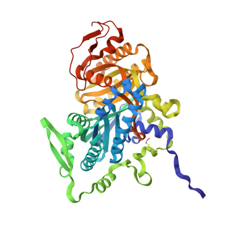
Crystal Structures of Human Soluble Adenylyl Cyclase Reveal Mechanisms of Catalysis and of its Activation Through Bicarbonate.

Kleinboelting, S.Diaz, A.Moniot, S.Van Den Heuvel, J.Weyand, M.Levin, L.R.Buck, J.Steegborn, C.

(2014) Proc Natl Acad Sci U S A 111: 3727

  • DOI: https://doi.org/10.1073/pnas.1322778111
  • Primary Citation of Related Structures:  
    4CLF, 4CLK, 4CLL, 4CLP, 4CLS, 4CLT, 4CLU, 4CLW, 4CLY, 4CLZ, 4CM0, 4CM2

  • PubMed Abstract: 

    cAMP is an evolutionary conserved, prototypic second messenger regulating numerous cellular functions. In mammals, cAMP is synthesized by one of 10 homologous adenylyl cyclases (ACs): nine transmembrane enzymes and one soluble AC (sAC). Among these, only sAC is directly activated by bicarbonate (HCO3(-)); it thereby serves as a cellular sensor for HCO3(-), carbon dioxide (CO2), and pH in physiological functions, such as sperm activation, aqueous humor formation, and metabolic regulation. Here, we describe crystal structures of human sAC catalytic domains in the apo state and in complex with substrate analog, products, and regulators. The activator HCO3(-) binds adjacent to Arg176, which acts as a switch that enables formation of the catalytic cation sites. An anionic inhibitor, 4,4'-diisothiocyanatostilbene-2,2'-disulfonic acid, inhibits sAC through binding to the active site entrance, which blocks HCO3(-) activation through steric hindrance and trapping of the Arg176 side chain. Finally, product complexes reveal small, local rearrangements that facilitate catalysis. Our results provide a molecular mechanism for sAC catalysis and cellular HCO3(-) sensing and a basis for targeting this system with drugs.


  • Organizational Affiliation

    Department of Biochemistry, University of Bayreuth, 95440 Bayreuth, Germany.


Macromolecules
Find similar proteins by:  (by identity cutoff)  |  3D Structure
Entity ID: 1
MoleculeChains Sequence LengthOrganismDetailsImage
ADENYLATE CYCLASE TYPE 10475Homo sapiensMutation(s): 0 
EC: 4.6.1.1
UniProt & NIH Common Fund Data Resources
Find proteins for Q96PN6 (Homo sapiens)
Explore Q96PN6 
Go to UniProtKB:  Q96PN6
PHAROS:  Q96PN6
GTEx:  ENSG00000143199 
Entity Groups  
Sequence Clusters30% Identity50% Identity70% Identity90% Identity95% Identity100% Identity
UniProt GroupQ96PN6
Sequence Annotations
Expand
  • Reference Sequence
Small Molecules
Modified Residues  1 Unique
IDChains TypeFormula2D DiagramParent
CME
Query on CME
A
L-PEPTIDE LINKINGC5 H11 N O3 S2CYS
Experimental Data & Validation

Experimental Data

  • Method: X-RAY DIFFRACTION
  • Resolution: 2.05 Å
  • R-Value Free: 0.197 
  • R-Value Work: 0.157 
  • R-Value Observed: 0.159 
  • Space Group: P 63
Unit Cell:
Length ( Å )Angle ( ˚ )
a = 100.17α = 90
b = 100.17β = 90
c = 97.49γ = 120
Software Package:
Software NamePurpose
REFMACrefinement
XDSdata reduction
XSCALEdata scaling
MOLREPphasing

Structure Validation

View Full Validation Report


Entry History 

Deposition Data

Revision History  (Full details and data files)

  • Version 1.0: 2014-03-05
    Type: Initial release
  • Version 1.1: 2014-03-26
    Changes: Database references
  • Version 1.2: 2019-04-03
    Changes: Data collection, Derived calculations, Other, Source and taxonomy
  • Version 1.3: 2023-12-20
    Changes: Data collection, Database references, Derived calculations, Other, Refinement description