Chronophin Dimerization is Required for Proper Positioning of its Substrate Specificity Loop.
Kestler, C., Knobloch, G., Tessmer, I., Jeanclos, E., Schindelin, H., Gohla, A.(2014) J Biol Chem 289: 3094
- PubMed: 24338687
- DOI: https://doi.org/10.1074/jbc.M113.536482
- Primary Citation of Related Structures:
4BX0, 4BX2, 4BX3 - PubMed Abstract:
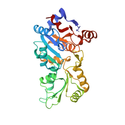
Mammalian phosphatases of the haloacid dehalogenase (HAD) superfamily have emerged as important regulators of physiology and disease. Many of these enzymes are stable homodimers; however, the role of their dimerization is largely unknown. Here, we explore the function of the obligatory homodimerization of chronophin, a mammalian HAD phosphatase known to dephosphorylate pyridoxal 5'-phosphate (PLP) and serine/threonine-phosphorylated proteins. The exchange of two residues in the murine chronophin homodimerization interface (chronophin(A194K,A195K)) yields a constitutive monomer both in vitro and in cells. The catalytic activity of monomeric chronophin toward PLP is strongly impaired. X-ray crystallographic studies of chronophin(A194K,A195K) revealed that dimer formation is essential for an intermolecular arginine-arginine-tryptophan stacking interaction that positions a critical histidine residue in the substrate specificity loop of chronophin for PLP coordination. Analysis of all available crystal structures of HAD hydrolases that are grouped together with chronophin in the C2a-type structural subfamily uncovered a highly conserved mode of dimerization that results in intermolecular contacts involving the substrate specificity loop. Our results explain how the dimerization of HAD hydrolases contributes to their catalytic efficiency and substrate specificity.
Organizational Affiliation:
From the Institute for Pharmacology and Toxicology, University of Würzburg, 97078 Würzburg, Germany and.