4BTE

DJ-1 Cu(I) complex


Experimental Data Snapshot

  • Method: X-RAY DIFFRACTION
  • Resolution: 1.38 Å
  • R-Value Free: 0.150 
  • R-Value Work: 0.118 
  • R-Value Observed: 0.120 

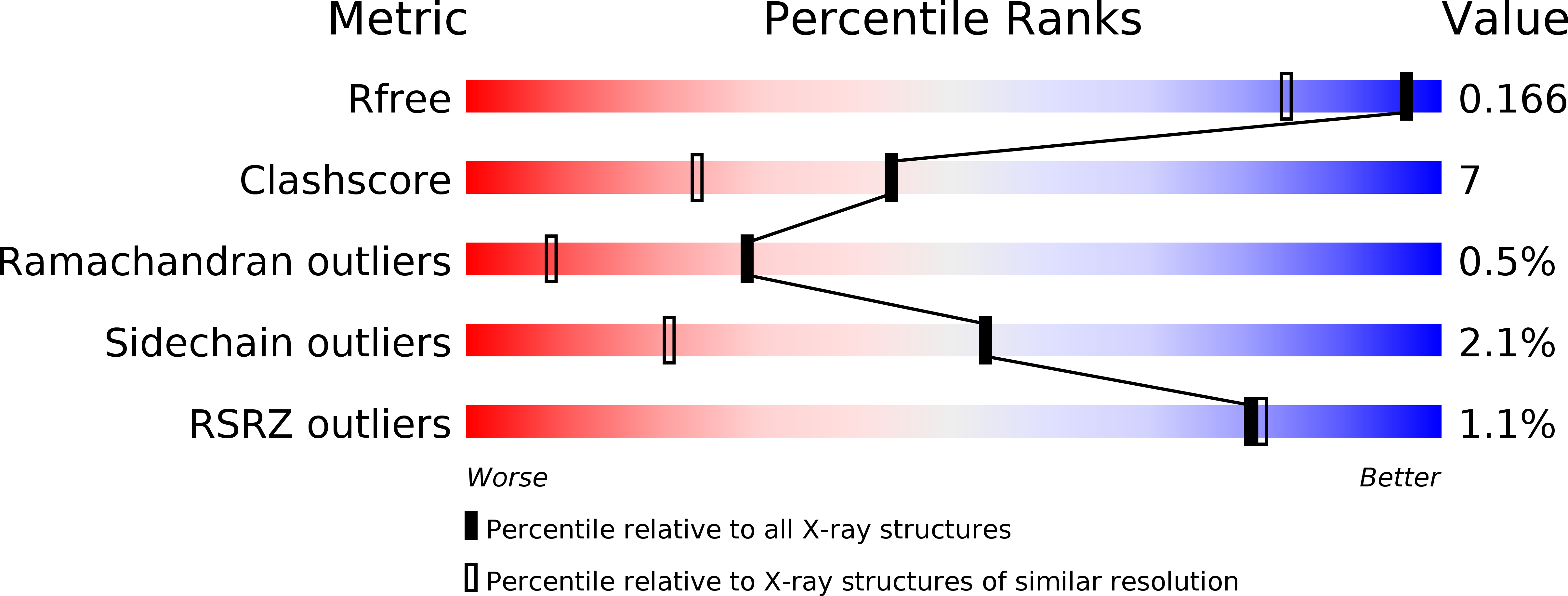
wwPDB Validation   3D Report Full Report


This is version 1.2 of the entry. See complete history


Literature

Structure of Cu(I)-Bound Dj-1 Reveals a Biscysteinate Metal Binding Site at the Homodimer Interface: Insights Into Mutational Inactivation of Dj-1 in Parkinsonism.

Puno, M.R.Patel, N.A.Moller, S.G.Robinson, C.V.Moody, P.C.E.Odell, M.

(2013) J Am Chem Soc 135: 15974

  • DOI: https://doi.org/10.1021/ja406010m
  • Primary Citation of Related Structures:  
    4BTE

  • PubMed Abstract: 

    The Parkinsonism-associated protein DJ-1 has been suggested to activate the Cu-Zn superoxide dismutase (SOD1) by providing its copper cofactor. The structural and chemical means by which DJ-1 could support this function is unknown. In this study, we characterize the molecular interaction of DJ-1 with Cu(I). Mass spectrometric analysis indicates binding of one Cu(I) ion per DJ-1 homodimer. The crystal structure of DJ-1 bound to Cu(I) confirms metal coordination through a docking accessible biscysteinate site formed by juxtaposed cysteine residues at the homodimer interface. Spectroscopy in crystallo validates the identity and oxidation state of the bound metal. The measured subfemtomolar dissociation constant (Kd = 6.41 × 10(-16) M) of DJ-1 for Cu(I) supports the physiological retention of the metal ion. Our results highlight the requirement of a stable homodimer for copper binding by DJ-1. Parkinsonism-linked mutations that weaken homodimer interactions will compromise this capability.


  • Organizational Affiliation

    Department of Molecular and Applied Biosciences, University of Westminster , 115 New Cavendish Street, London W1W 6UW, United Kingdom.


Macromolecules
Find similar proteins by:  (by identity cutoff)  |  3D Structure
Entity ID: 1
MoleculeChains Sequence LengthOrganismDetailsImage
PROTEIN DJ-1189Homo sapiensMutation(s): 0 
EC: 3.4
UniProt & NIH Common Fund Data Resources
Find proteins for Q99497 (Homo sapiens)
Explore Q99497 
Go to UniProtKB:  Q99497
PHAROS:  Q99497
GTEx:  ENSG00000116288 
Entity Groups  
Sequence Clusters30% Identity50% Identity70% Identity90% Identity95% Identity100% Identity
UniProt GroupQ99497
Sequence Annotations
Expand
  • Reference Sequence
Small Molecules
Ligands 1 Unique
IDChains Name / Formula / InChI Key2D Diagram3D Interactions
CU1
Query on CU1

Download Ideal Coordinates CCD File 
B [auth A]COPPER (I) ION
Cu
VMQMZMRVKUZKQL-UHFFFAOYSA-N
Experimental Data & Validation

Experimental Data

  • Method: X-RAY DIFFRACTION
  • Resolution: 1.38 Å
  • R-Value Free: 0.150 
  • R-Value Work: 0.118 
  • R-Value Observed: 0.120 
  • Space Group: P 31 2 1
Unit Cell:
Length ( Å )Angle ( ˚ )
a = 75.257α = 90
b = 75.257β = 90
c = 75.354γ = 120
Software Package:
Software NamePurpose
REFMACrefinement
XDSdata reduction
SCALAdata scaling
PHASERphasing

Structure Validation

View Full Validation Report


Entry History 

Deposition Data

Revision History  (Full details and data files)

  • Version 1.0: 2013-11-06
    Type: Initial release
  • Version 1.1: 2013-12-25
    Changes: Database references
  • Version 1.2: 2023-12-20
    Changes: Data collection, Database references, Derived calculations, Other, Refinement description