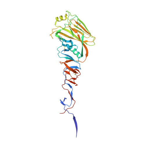
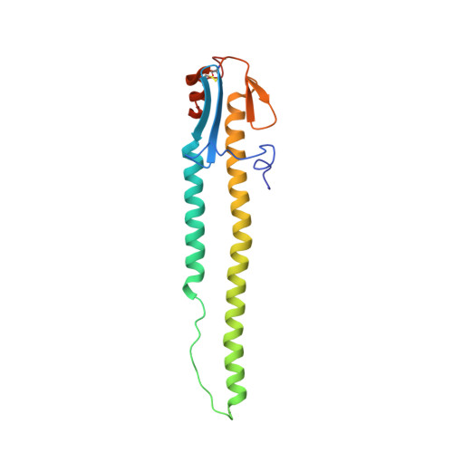
4BGY

H5 (VN1194) Influenza Virus Haemagglutinin in Complex with Avian Receptor Analogue 3'-SLN


Experimental Data Snapshot

  • Method: X-RAY DIFFRACTION
  • Resolution: 2.68 Å
  • R-Value Free: 0.248 
  • R-Value Work: 0.202 
  • R-Value Observed: 0.204 

wwPDB Validation   3D Report Full Report


This is version 2.0 of the entry. See complete history


Literature

Receptor Binding by a Ferret-Transmissible H5 Avian Influenza Virus.

Xiong, X.Coombs, P.R Martin, S.Liu, J.Xiao, H.Mccauley, J.W.Locher, K.Walker, P.A.Collins, P.J.Kawaoka, Y.Skehel, J.J.Gamblin, S.J.

(2013) Nature 497: 392

  • DOI: https://doi.org/10.1038/nature12144
  • Primary Citation of Related Structures:  
    4BGW, 4BGX, 4BGY, 4BGZ, 4BH0, 4BH1, 4BH2, 4BH3, 4BH4

  • PubMed Abstract: 

    Cell-surface-receptor binding by influenza viruses is a key determinant of their transmissibility, both from avian and animal species to humans as well as from human to human. Highly pathogenic avian H5N1 viruses that are a threat to public health have been observed to acquire affinity for human receptors, and transmissible-mutant-selection experiments have identified a virus that is transmissible in ferrets, the generally accepted experimental model for influenza in humans. Here, our quantitative biophysical measurements of the receptor-binding properties of haemagglutinin (HA) from the transmissible mutant indicate a small increase in affinity for human receptor and a marked decrease in affinity for avian receptor. From analysis of virus and HA binding data we have derived an algorithm that predicts virus avidity from the affinity of individual HA-receptor interactions. It reveals that the transmissible-mutant virus has a 200-fold preference for binding human over avian receptors. The crystal structure of the transmissible-mutant HA in complex with receptor analogues shows that it has acquired the ability to bind human receptor in the same folded-back conformation as seen for HA from the 1918, 1957 (ref. 4), 1968 (ref. 5) and 2009 (ref. 6) pandemic viruses. This binding mode is substantially different from that by which non-transmissible wild-type H5 virus HA binds human receptor. The structure of the complex also explains how the change in preference from avian to human receptors arises from the Gln226Leu substitution, which facilitates binding to human receptor but restricts binding to avian receptor. Both features probably contribute to the acquisition of transmissibility by this mutant virus.


  • Organizational Affiliation

    MRC National Institute for Medical Research, The Ridgeway, Mill Hill, London NW7 1AA, UK.


Macromolecules
Find similar proteins by:  (by identity cutoff)  |  3D Structure
Entity ID: 1
MoleculeChains Sequence LengthOrganismDetailsImage
HEMAGGLUTININ326Influenza A virus (A/Vietnam/1194/2004(H5N1))Mutation(s): 0 
UniProt
Find proteins for Q6DQ34 (Influenza A virus)
Explore Q6DQ34 
Go to UniProtKB:  Q6DQ34
Entity Groups  
Sequence Clusters30% Identity50% Identity70% Identity90% Identity95% Identity100% Identity
UniProt GroupQ6DQ34
Sequence Annotations
Expand
  • Reference Sequence
Find similar proteins by:  (by identity cutoff)  |  3D Structure
Entity ID: 2
MoleculeChains Sequence LengthOrganismDetailsImage
HEMAGGLUTININ166Influenza A virus (A/Vietnam/1194/2004(H5N1))Mutation(s): 0 
UniProt
Find proteins for Q6DQ34 (Influenza A virus)
Explore Q6DQ34 
Go to UniProtKB:  Q6DQ34
Entity Groups  
Sequence Clusters30% Identity50% Identity70% Identity90% Identity95% Identity100% Identity
UniProt GroupQ6DQ34
Sequence Annotations
Expand
  • Reference Sequence
Oligosaccharides

Help

Entity ID: 3
MoleculeChains Length2D Diagram Glycosylation3D Interactions
2-acetamido-2-deoxy-beta-D-glucopyranose-(1-4)-2-acetamido-2-deoxy-beta-D-glucopyranose
C, D, F
2N-Glycosylation
Glycosylation Resources
GlyTouCan:  G42666HT
GlyCosmos:  G42666HT
GlyGen:  G42666HT
Entity ID: 4
MoleculeChains Length2D Diagram Glycosylation3D Interactions
N-acetyl-alpha-neuraminic acid-(2-3)-beta-D-galactopyranose-(1-4)-2-acetamido-2-deoxy-beta-D-glucopyranose
E
3N/A
Glycosylation Resources
GlyTouCan:  G00065MO
GlyCosmos:  G00065MO
GlyGen:  G00065MO
Small Molecules
Ligands 1 Unique
IDChains Name / Formula / InChI Key2D Diagram3D Interactions
EPE
Query on EPE

Download Ideal Coordinates CCD File 
G [auth B]4-(2-HYDROXYETHYL)-1-PIPERAZINE ETHANESULFONIC ACID
C8 H18 N2 O4 S
JKMHFZQWWAIEOD-UHFFFAOYSA-N
Biologically Interesting Molecules (External Reference) 1 Unique
Experimental Data & Validation

Experimental Data

  • Method: X-RAY DIFFRACTION
  • Resolution: 2.68 Å
  • R-Value Free: 0.248 
  • R-Value Work: 0.202 
  • R-Value Observed: 0.204 
  • Space Group: H 3 2
Unit Cell:
Length ( Å )Angle ( ˚ )
a = 101.403α = 90
b = 101.403β = 90
c = 449.997γ = 120
Software Package:
Software NamePurpose
REFMACrefinement
MOSFLMdata reduction
SCALAdata scaling
PHASERphasing

Structure Validation

View Full Validation Report


Entry History 

Deposition Data

Revision History  (Full details and data files)

  • Version 1.0: 2013-04-24
    Type: Initial release
  • Version 1.1: 2013-05-08
    Changes: Database references, Refinement description
  • Version 1.2: 2013-05-15
    Changes: Database references
  • Version 1.3: 2013-05-22
    Changes: Database references
  • Version 2.0: 2020-07-29
    Type: Remediation
    Reason: Carbohydrate remediation
    Changes: Advisory, Atomic model, Data collection, Derived calculations, Other, Structure summary