4BEJ

Nucleotide-free Dynamin 1-like protein (DNM1L, DRP1, DLP1)


Experimental Data Snapshot

  • Method: X-RAY DIFFRACTION
  • Resolution: 3.48 Å
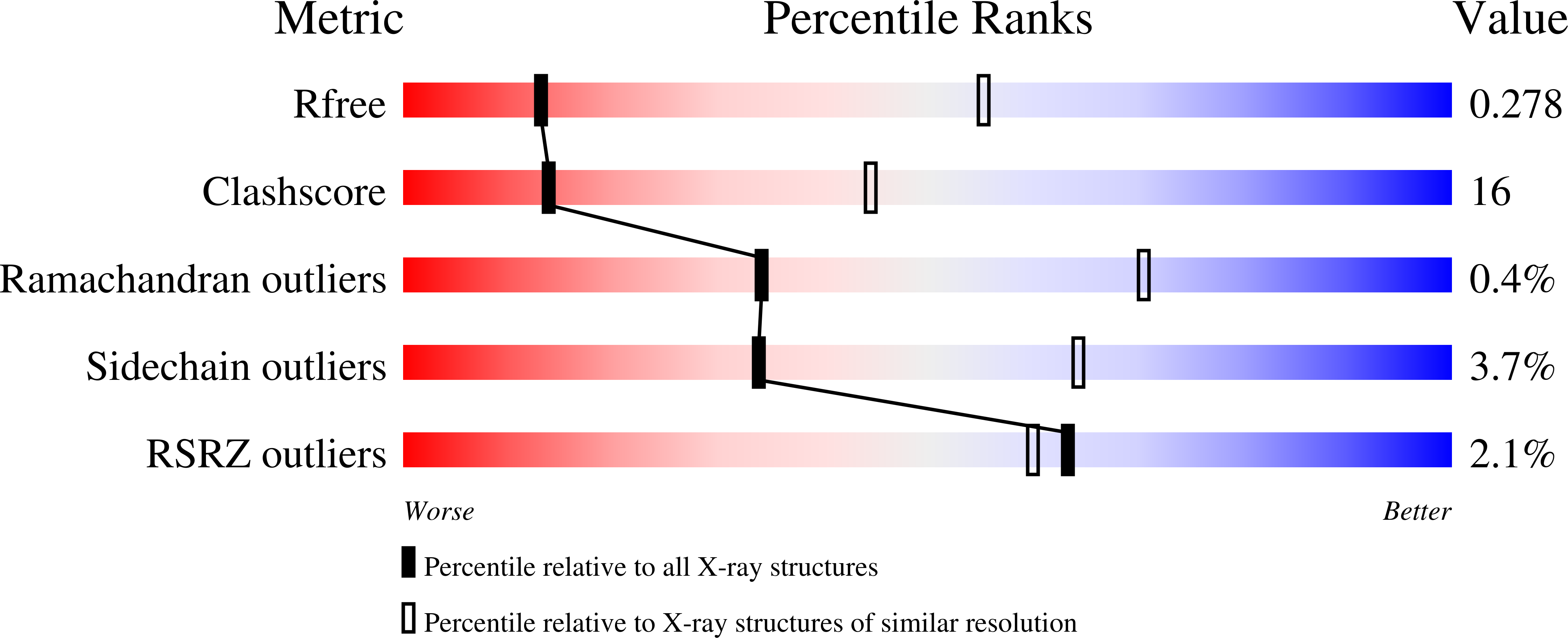
  • R-Value Free: 0.276 
  • R-Value Work: 0.251 
  • R-Value Observed: 0.252 

wwPDB Validation   3D Report Full Report


This is version 1.2 of the entry. See complete history


Literature

Structural Insights Into Oligomerization and Mitochondrial Remodelling of Dynamin 1-Like Protein.

Frohlich, C.Grabiger, S.Schwefel, D.Faelber, K.Rosenbaum, E.Mears, J.Rocks, O.Daumke, O.

(2013) EMBO J 32: 1280

  • DOI: https://doi.org/10.1038/emboj.2013.74
  • Primary Citation of Related Structures:  
    4BEJ

  • PubMed Abstract: 

    Dynamin 1-like protein (DNM1L) mediates fission of mitochondria and peroxisomes, and dysfunction of DNM1L has been implicated in several neurological disorders. To study the molecular basis of mitochondrial remodelling, we determined the crystal structure of DNM1L that is comprised of a G domain, a bundle signalling element and a stalk. DNM1L assembled via a central stalk interface, and mutations in this interface disrupted dimerization and interfered with membrane binding and mitochondrial targeting. Two sequence stretches at the tip of the stalk were shown to be required for ordered assembly of DNM1L on membranes and its function in mitochondrial fission. In the crystals, DNM1L dimers further assembled via a second, previously undescribed, stalk interface to form a linear filament. Mutations in this interface interfered with liposome tubulation and mitochondrial remodelling. Based on these results and electron microscopy reconstructions, we propose an oligomerization mode for DNM1L which differs from that of dynamin and might be adapted to the remodelling of mitochondria.


  • Organizational Affiliation

    Crystallography, Max-Delbrück-Center for Molecular Medicine, Berlin, Germany.


Macromolecules
Find similar proteins by:  (by identity cutoff)  |  3D Structure
Entity ID: 1
MoleculeChains Sequence LengthOrganismDetailsImage
DYNAMIN 1-LIKE PROTEIN
A, B, C, D
617Homo sapiensMutation(s): 4 
EC: 3.6.5.5
UniProt & NIH Common Fund Data Resources
Find proteins for O00429 (Homo sapiens)
Explore O00429 
Go to UniProtKB:  O00429
PHAROS:  O00429
GTEx:  ENSG00000087470 
Entity Groups  
Sequence Clusters30% Identity50% Identity70% Identity90% Identity95% Identity100% Identity
UniProt GroupO00429
Sequence Annotations
Expand
  • Reference Sequence
Experimental Data & Validation

Experimental Data

  • Method: X-RAY DIFFRACTION
  • Resolution: 3.48 Å
  • R-Value Free: 0.276 
  • R-Value Work: 0.251 
  • R-Value Observed: 0.252 
  • Space Group: P 1 2 1
Unit Cell:
Length ( Å )Angle ( ˚ )
a = 101.47α = 90
b = 80.77β = 93.45
c = 208.272γ = 90
Software Package:
Software NamePurpose
Cootmodel building
XDSdata scaling
XDSphasing
CCP4phasing
PHENIXphasing
Cootphasing
PHENIXrefinement

Structure Validation

View Full Validation Report


Entry History 

Deposition Data

Revision History  (Full details and data files)

  • Version 1.0: 2013-04-24
    Type: Initial release
  • Version 1.1: 2013-05-22
    Changes: Database references
  • Version 1.2: 2023-12-20
    Changes: Data collection, Database references, Other, Refinement description