4AXG

Structure of eIF4E-Cup complex


Experimental Data Snapshot

  • Method: X-RAY DIFFRACTION
  • Resolution: 2.80 Å
  • R-Value Free: 0.244 
  • R-Value Work: 0.228 
  • R-Value Observed: 0.229 

wwPDB Validation   3D Report Full Report


This is version 1.1 of the entry. See complete history


Literature

Crystal Structure of a Minimal Eif4E-Cup Complex Revelas a General Mechanism of Eif4E Regulation in Translational Repression

Kinkelin, K.Veith, K.Gruenwald, M.Bono, F.

(2012) RNA 18: 1624

  • DOI: https://doi.org/10.1261/rna.033639.112
  • Primary Citation of Related Structures:  
    4AXG

  • PubMed Abstract: 

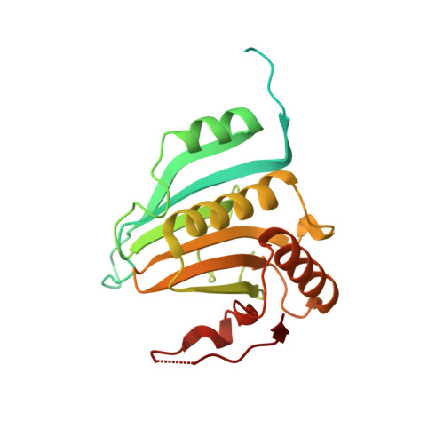
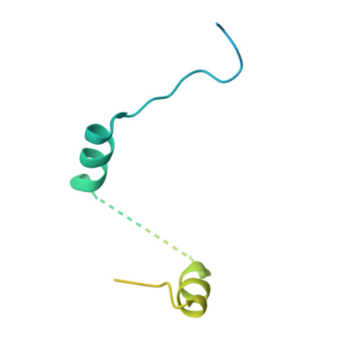
    Cup is an eIF4E-binding protein (4E-BP) that plays a central role in translational regulation of localized mRNAs during early Drosophila development. In particular, Cup is required for repressing translation of the maternally contributed oskar, nanos, and gurken mRNAs, all of which are essential for embryonic body axis determination. Here, we present the 2.8 Å resolution crystal structure of a minimal eIF4E-Cup assembly, consisting of the interacting regions of the two proteins. In the structure, two separate segments of Cup contact two orthogonal faces of eIF4E. The eIF4E-binding consensus motif of Cup (YXXXXLΦ) binds the convex side of eIF4E similarly to the consensus of other eIF4E-binding proteins, such as 4E-BPs and eIF4G. The second, noncanonical, eIF4E-binding site of Cup binds laterally and perpendicularly to the eIF4E β-sheet. Mutations of Cup at this binding site were shown to reduce binding to eIF4E and to promote the destabilization of the associated mRNA. Comparison with the binding mode of eIF4G to eIF4E suggests that Cup and eIF4G binding would be mutually exclusive at both binding sites. This shows how a common molecular surface of eIF4E might recognize different proteins acting at different times in the same pathway. The structure provides insight into the mechanism by which Cup disrupts eIF4E-eIF4G interaction and has broader implications for understanding the role of 4E-BPs in translational regulation.


  • Organizational Affiliation

    Max-Planck-Institute for Developmental Biology, 71076 Tübingen, Germany.


Macromolecules
Find similar proteins by:  (by identity cutoff)  |  3D Structure
Entity ID: 1
MoleculeChains Sequence LengthOrganismDetailsImage
EUKARYOTIC TRANSLATION INITIATION FACTOR 4E
A, B
248Drosophila melanogasterMutation(s): 0 
UniProt
Find proteins for P48598 (Drosophila melanogaster)
Explore P48598 
Go to UniProtKB:  P48598
Entity Groups  
Sequence Clusters30% Identity50% Identity70% Identity90% Identity95% Identity100% Identity
UniProt GroupP48598
Sequence Annotations
Expand
  • Reference Sequence
Find similar proteins by:  (by identity cutoff)  |  3D Structure
Entity ID: 2
MoleculeChains Sequence LengthOrganismDetailsImage
PROTEIN CUP
C, D
130Drosophila melanogasterMutation(s): 0 
UniProt
Find proteins for Q9VMA3 (Drosophila melanogaster)
Explore Q9VMA3 
Go to UniProtKB:  Q9VMA3
Entity Groups  
Sequence Clusters30% Identity50% Identity70% Identity90% Identity95% Identity100% Identity
UniProt GroupQ9VMA3
Sequence Annotations
Expand
  • Reference Sequence
Experimental Data & Validation

Experimental Data

  • Method: X-RAY DIFFRACTION
  • Resolution: 2.80 Å
  • R-Value Free: 0.244 
  • R-Value Work: 0.228 
  • R-Value Observed: 0.229 
  • Space Group: P 61 2 2
Unit Cell:
Length ( Å )Angle ( ˚ )
a = 142.58α = 90
b = 142.58β = 90
c = 108.05γ = 120
Software Package:
Software NamePurpose
PHENIXrefinement
XDSdata reduction
XDSdata scaling
PHASERphasing

Structure Validation

View Full Validation Report


Entry History 

Deposition Data

Revision History  (Full details and data files)

  • Version 1.0: 2012-08-29
    Type: Initial release
  • Version 1.1: 2023-12-20
    Changes: Data collection, Database references, Other, Refinement description