4AWE

The Crystal Structure of Chrysonilia sitophila endo-beta-D-1,4- mannanase


Experimental Data Snapshot

  • Method: X-RAY DIFFRACTION
  • Resolution: 1.40 Å
  • R-Value Free: 0.202 
  • R-Value Work: 0.161 
  • R-Value Observed: 0.163 

wwPDB Validation   3D Report Full Report


Ligand Structure Quality Assessment 


This is version 2.1 of the entry. See complete history


Literature

Endo-Beta-D-1,4-Mannanase from Chrysonilia Sitophila Displays a Novel Loop Arrangement for Substrate Selectivity

Goncalves, A.M.D.Silva, C.S.Madeira, T.I.Coelho, R.De Sanctis, D.San Romao, M.V.Bento, I.

(2012) Acta Crystallogr D Biol Crystallogr 68: 1468

  • DOI: https://doi.org/10.1107/S0907444912034646
  • Primary Citation of Related Structures:  
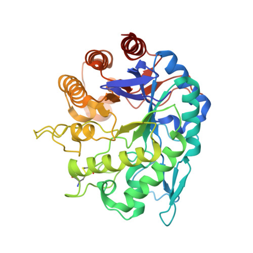
    4AWE

  • PubMed Abstract: 

    The crystal structure of wild-type endo-β-D-1,4-mannanase (EC 3.2.1.78) from the ascomycete Chrysonilia sitophila (CsMan5) has been solved at 1.40 Å resolution. The enzyme isolated directly from the source shows mixed activity as both an endo-glucanase and an endo-mannanase. CsMan5 adopts the (β/α)(8)-barrel fold that is well conserved within the GH5 family and has highest sequence and structural homology to the GH5 endo-mannanases. Superimposition with proteins of this family shows a unique structural arrangement of three surface loops of CsMan5 that stretch over the active centre, promoting an altered topography of the binding cleft. The most relevant feature results from the repositioning of a long loop at the extremity of the binding cleft, resulting in a shortened glycone-binding region with two subsites. The other two extended loops flanking the binding groove produce a narrower cleft compared with the wide architecture observed in GH5 homologues. Two aglycone subsites (+1 and +2) are identified and a nonconserved tryptophan (Trp271) at the +1 subsite may offer steric hindrance. Taken together, these findings suggest that the discrimination of mannan substrates is achieved through modified loop length and structure.


  • Organizational Affiliation

    Macromolecular Crystallography Unit, Instituto de Tecnologia Química e Biológica, Universidade Nova de Lisboa, Avenida da República, 2780-157 Oeiras, Portugal.


Macromolecules
Find similar proteins by:  (by identity cutoff)  |  3D Structure
Entity ID: 1
MoleculeChains Sequence LengthOrganismDetailsImage
ENDO-BETA-D-1,4-MANNANASE387Neurospora sitophilaMutation(s): 0 
EC: 3.2.1.78
UniProt
Find proteins for I4IY26 (Neurospora sitophila)
Explore I4IY26 
Go to UniProtKB:  I4IY26
Entity Groups  
Sequence Clusters30% Identity50% Identity70% Identity90% Identity95% Identity100% Identity
UniProt GroupI4IY26
Sequence Annotations
Expand
  • Reference Sequence
Oligosaccharides

Help

Entity ID: 2
MoleculeChains Length2D Diagram Glycosylation3D Interactions
2-acetamido-2-deoxy-beta-D-glucopyranose-(1-4)-2-acetamido-2-deoxy-beta-D-glucopyranose
B
2N-Glycosylation
Glycosylation Resources
GlyTouCan:  G42666HT
GlyCosmos:  G42666HT
GlyGen:  G42666HT
Experimental Data & Validation

Experimental Data

  • Method: X-RAY DIFFRACTION
  • Resolution: 1.40 Å
  • R-Value Free: 0.202 
  • R-Value Work: 0.161 
  • R-Value Observed: 0.163 
  • Space Group: P 21 21 21
Unit Cell:
Length ( Å )Angle ( ˚ )
a = 57.36α = 90
b = 78.94β = 90
c = 83.86γ = 90
Software Package:
Software NamePurpose
REFMACrefinement
MOSFLMdata reduction
SCALAdata scaling
PHASERphasing

Structure Validation

View Full Validation Report


Ligand Structure Quality Assessment 


Entry History 

Deposition Data

Revision History  (Full details and data files)

  • Version 1.0: 2012-10-31
    Type: Initial release
  • Version 1.1: 2012-11-07
    Changes: Database references
  • Version 2.0: 2020-07-29
    Type: Remediation
    Reason: Carbohydrate remediation
    Changes: Advisory, Atomic model, Data collection, Derived calculations, Other, Structure summary
  • Version 2.1: 2023-12-20
    Changes: Data collection, Database references, Refinement description, Structure summary