4AKJ

Ligand controlled assembly of hexamers, dihexamers, and linear multihexamer structures by an engineered acylated insulin


Experimental Data Snapshot

  • Method: X-RAY DIFFRACTION
  • Resolution: 2.01 Å
  • R-Value Free: 0.229 
  • R-Value Work: 0.196 
  • R-Value Observed: 0.198 

wwPDB Validation   3D Report Full Report


Ligand Structure Quality Assessment 


This is version 1.3 of the entry. See complete history


Literature

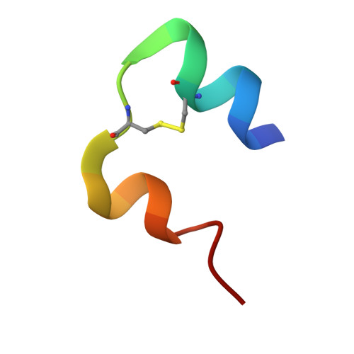
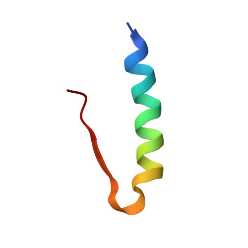
Ligand Controlled Assembly of Hexamers, Dihexamers, and Linear Multihexamer Structures by the Engineered Acylated Insulin Degludec.

Steensgaard, D.B.Schluckebier, G.Strauss, H.M.Norrman, M.Thomsen, J.K.Friderichsen, A.V.Havelund, S.Jonassen, I.

(2013) Biochemistry 52: 295

  • DOI: https://doi.org/10.1021/bi3008609
  • Primary Citation of Related Structures:  
    4AJX, 4AJZ, 4AK0, 4AKJ

  • PubMed Abstract: 

    Insulin degludec, an engineered acylated insulin, was recently reported to form a soluble depot after subcutaneous injection with a subsequent slow release of insulin and an ultralong glucose-lowering effect in excess of 40 h in humans. We describe the structure, ligand binding properties, and self-assemblies of insulin degludec using orthogonal structural methods. The protein fold adopted by insulin degludec is very similar to that of human insulin. Hexamers in the R(6) state similar to those of human insulin are observed for insulin degludec in the presence of zinc and resorcinol. However, under conditions comparable to the pharmaceutical formulation comprising zinc and phenol, insulin degludec forms finite dihexamers that are composed of hexamers in the T(3)R(3) state that interact to form an R(3)T(3)-T(3)R(3) structure. When the phenolic ligand is depleted and the solvent condition thereby mimics that of the injection site, the quaternary structure changes from dihexamers to a supramolecular structure composed of linear arrays of hundreds of hexamers in the T(6) state and an average molar mass, M(0), of 59.7 × 10(3) kg/mol. This novel concept of self-assemblies of insulin controlled by zinc and phenol provides the basis for the slow action profile of insulin degludec. To the best of our knowledge, this report for the first time describes a tight linkage between quaternary insulin structures of hexamers, dihexamers, and multihexamers and their allosteric state and its origin in the inherent propensity of the insulin hexamer for allosteric half-site reactivity.


  • Organizational Affiliation

    Diabetes Protein Engineering, Novo Nordisk A/S , Novo Nordisk Park, 2760 Maaloev, Denmark. dbs@novonordisk.com


Macromolecules

Find similar proteins by:  Sequence   |   3D Structure  

Entity ID: 1
MoleculeChains Sequence LengthOrganismDetailsImage
INSULIN A CHAIN
A, C
21Homo sapiensMutation(s): 0 
UniProt & NIH Common Fund Data Resources
Find proteins for P01308 (Homo sapiens)
Explore P01308 
Go to UniProtKB:  P01308
PHAROS:  P01308
GTEx:  ENSG00000254647 
Entity Groups  
Sequence Clusters30% Identity50% Identity70% Identity90% Identity95% Identity100% Identity
UniProt GroupP01308
Sequence Annotations
Expand
  • Reference Sequence
Find similar proteins by:  (by identity cutoff)  |  3D Structure
Entity ID: 2
MoleculeChains Sequence LengthOrganismDetailsImage
INSULIN B CHAIN
B, D
29Homo sapiensMutation(s): 0 
UniProt & NIH Common Fund Data Resources
Find proteins for P01308 (Homo sapiens)
Explore P01308 
Go to UniProtKB:  P01308
PHAROS:  P01308
GTEx:  ENSG00000254647 
Entity Groups  
Sequence Clusters30% Identity50% Identity70% Identity90% Identity95% Identity100% Identity
UniProt GroupP01308
Sequence Annotations
Expand
  • Reference Sequence
Small Molecules
Ligands 4 Unique
IDChains Name / Formula / InChI Key2D Diagram3D Interactions
16E
Query on 16E

Download Ideal Coordinates CCD File 
F [auth B]N-(16-Carboxyhexadecanoyl)-L-glutamic acid
C21 H37 N O7
SGIVALYPXQHYAY-KRWDZBQOSA-N
IPH
Query on IPH

Download Ideal Coordinates CCD File 
E [auth A],
I [auth C]
PHENOL
C6 H6 O
ISWSIDIOOBJBQZ-UHFFFAOYSA-N
ZN
Query on ZN

Download Ideal Coordinates CCD File 
G [auth B],
J [auth D]
ZINC ION
Zn
PTFCDOFLOPIGGS-UHFFFAOYSA-N
CL
Query on CL

Download Ideal Coordinates CCD File 
H [auth B],
K [auth D]
CHLORIDE ION
Cl
VEXZGXHMUGYJMC-UHFFFAOYSA-M
Experimental Data & Validation

Experimental Data

  • Method: X-RAY DIFFRACTION
  • Resolution: 2.01 Å
  • R-Value Free: 0.229 
  • R-Value Work: 0.196 
  • R-Value Observed: 0.198 
  • Space Group: H 3
Unit Cell:
Length ( Å )Angle ( ˚ )
a = 78.717α = 90
b = 78.717β = 90
c = 40.551γ = 120
Software Package:
Software NamePurpose
REFMACrefinement
HKL-2000data reduction
SCALEPACKdata scaling
MOLREPphasing

Structure Validation

View Full Validation Report


Ligand Structure Quality Assessment 


Entry History 

Deposition Data

Revision History  (Full details and data files)

  • Version 1.0: 2013-01-09
    Type: Initial release
  • Version 1.1: 2013-01-30
    Changes: Database references
  • Version 1.2: 2017-07-05
    Changes: Data collection
  • Version 1.3: 2023-12-20
    Changes: Data collection, Database references, Derived calculations, Other, Refinement description