Exploring the Chemical Space Around 8-Mercaptoguanine as a Route to New Inhibitors of the Folate Biosynthesis Enzyme Hppk.
Chhabra, S., Barlow, N., Dolezal, O., Hattarki, M.K., Newman, J., Peat, T.S., Graham, B., Swarbrick, J.D.(2013) PLoS One 8: 59535
- PubMed: 23565155
- DOI: https://doi.org/10.1371/journal.pone.0059535
- Primary Citation of Related Structures:
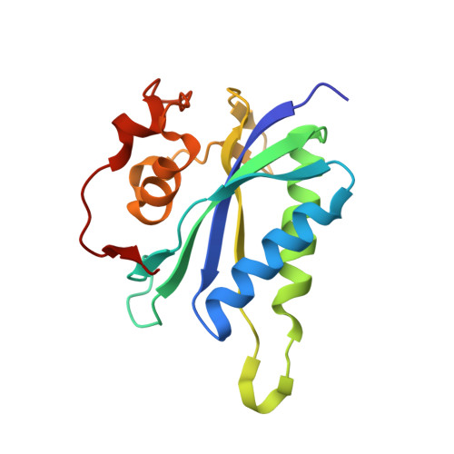
4AD6 - PubMed Abstract:
As the second essential enzyme of the folate biosynthetic pathway, the potential antimicrobial target, HPPK (6-hydroxymethyl-7,8-dihydropterin pyrophosphokinase), catalyzes the Mg(2+-)dependant transfer of pyrophosphate from the cofactor (ATP) to the substrate, 6-hydroxymethyl-7,8-dihydropterin. Recently, we showed that 8-mercaptoguanine (8-MG) bound at the substrate site (KD ∼13 µM), inhibited the S. aureus enzyme (SaHPPK) (IC50 ∼ 41 µM), and determined the structure of the SaHPPK/8-MG complex. Here we present the synthesis of a series of guanine derivatives, together with their HPPK binding affinities, as determined by SPR and ITC analysis. The binding mode of the most potent was investigated using 2D NMR spectroscopy and X-ray crystallography. The results indicate, firstly, that the SH group of 8-MG makes a significant contribution to the free energy of binding. Secondly, direct N(9) substitution, or tautomerization arising from N(7) substitution in some cases, leads to a dramatic reduction in affinity due to loss of a critical N(9)-H···Val46 hydrogen bond, combined with the limited space available around the N(9) position. The water-filled pocket under the N(7) position is significantly more tolerant of substitution, with a hydroxyl ethyl 8-MG derivative attached to N(7) (compound 21a) exhibiting an affinity for the apo enzyme comparable to the parent compound (KD ∼ 12 µM). In contrast to 8-MG, however, 21a displays competitive binding with the ATP cofactor, as judged by NMR and SPR analysis. The 1.85 Å X-ray structure of the SaHPPK/21a complex confirms that extension from the N(7) position towards the Mg(2+)-binding site, which affords the only tractable route out from the pterin-binding pocket. Promising strategies for the creation of more potent binders might therefore include the introduction of groups capable of interacting with the Mg(2+) centres or Mg(2+)-binding residues, as well as the development of bitopic inhibitors featuring 8-MG linked to a moiety targeting the ATP cofactor binding site.
Organizational Affiliation:
Medicinal Chemistry, Monash Institute of Pharmaceutical Sciences, Monash University, Parkville, Australia.