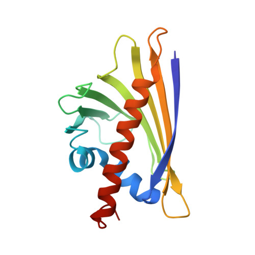
Crystallographically Mapped Ligand Binding Differs in High and Low Ige Binding Isoforms of Birch Pollen Allergen Bet V 1.
Kofler, S., Asam, C., Eckhard, U., Wallner, M., Ferreira, F., Brandstetter, H.(2012) J Mol Biol 422: 109
- PubMed: 22634284
- DOI: https://doi.org/10.1016/j.jmb.2012.05.016
- Primary Citation of Related Structures:
4A80, 4A81, 4A83, 4A84, 4A85, 4A86, 4A87, 4A88, 4A8G, 4A8U, 4A8V - PubMed Abstract:
The ability of pathogenesis-related proteins of family 10 to bind a broad spectrum of ligands is considered to play a key role for their physiological and pathological functions. In particular, Bet v 1, an archetypical allergen from birch pollen, is described as a highly promiscuous ligand acceptor. However, the detailed recognition mechanisms, including specificity factors discriminating binding properties of naturally occurring Bet v 1 variants, are poorly understood. Here, we report crystal structures of Bet v 1 variants in complex with an array of ligands at a resolution of up to 1.2 Å. Residue 30 within the hydrophobic pocket not only discriminates in high and low IgE binding Bet v 1 isoforms but also induces a drastic change in the binding mode of the model ligand deoxycholate. Ternary crystal structure complexes of Bet v 1 with several ligands together with the fluorogenic reporter 1-anilino-8-naphthalene sulfonate explain anomalous fluorescence binding curves obtained from 1-anilino-8-naphthalene sulfonate displacement assays. The structures reveal key interaction residues such as Tyr83 and rationalize both the binding specificity and promiscuity of the so-called hydrophobic pocket in Bet v 1. The intermolecular interactions of Bet v 1 reveal an unexpected complexity that will be indispensable to fully understand its roles within the physiological and allergenic context.
Organizational Affiliation:
Structural Biology Group, Department of Molecular Biology, University of Salzburg, A-5020 Salzburg, Austria.