


Structural Studies of Intermediates Along the Cyclization Pathway of Aplysia Adp-Ribosyl Cyclase.
Kotaka, M., Graeff, R., Chen, Z., Zhang, L.H., Lee, H.C., Hao, Q.To be published.
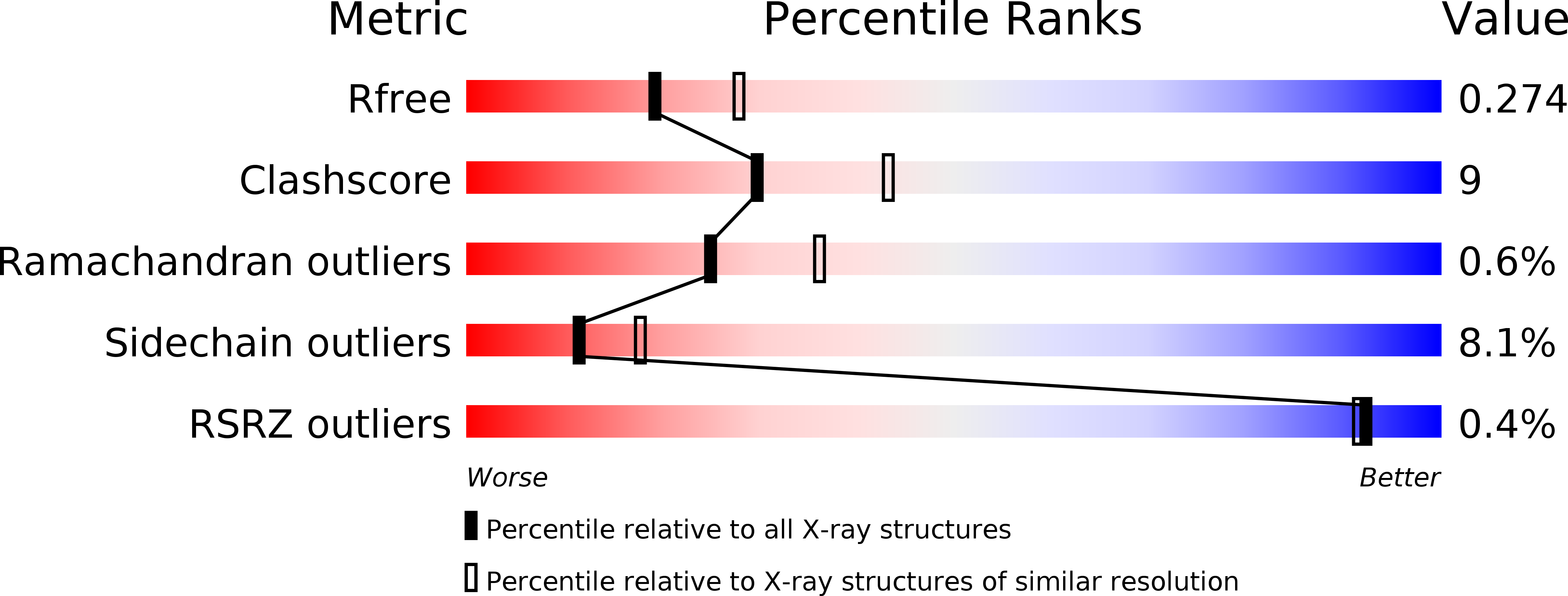
Experimental Data Snapshot
Entity ID: 1 | |||||
|---|---|---|---|---|---|
| Molecule | Chains | Sequence Length | Organism | Details | Image |
| ADP-RIBOSYL CYCLASE | 260 | Aplysia californica | Mutation(s): 0 EC: 3.2.2.5 | 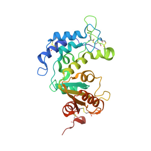 | |
UniProt | |||||
Find proteins for P29241 (Aplysia californica) Explore P29241 Go to UniProtKB: P29241 | |||||
Entity Groups | |||||
| Sequence Clusters | 30% Identity50% Identity70% Identity90% Identity95% Identity100% Identity | ||||
| UniProt Group | P29241 | ||||
Sequence AnnotationsExpand | |||||
| |||||
| Ligands 2 Unique | |||||
|---|---|---|---|---|---|
| ID | Chains | Name / Formula / InChI Key | 2D Diagram | 3D Interactions | |
| AV1 Query on AV1 | I [auth A] J [auth B] K [auth C] M [auth E] N [auth F] | [(2R,3S,4R,5R)-5-(6-amino-8-bromo-9H-purin-9-yl)-3,4-dihydroxytetrahydrofuran-2-yl]methyl [(2R,3S,4S)-3,4-dihydroxytetrahydrofuran-2-yl]methyl dihydrogen diphosphate C15 H22 Br N5 O13 P2 DTBPYRLOWSYQCZ-ZVIMGNLFSA-L |  | ||
| CV1 Query on CV1 | L [auth D] | (2R,3R,4S,5R,13R,14S,15R,16R)-24-amino-18-bromo-3,4,14,15-tetrahydroxy-7,9,11,25,26-pentaoxa-17,19,22-triaza-1-azonia-8
,10-diphosphapentacyclo[18.3.1.1^2,5^.1^13,16^.0^17,21^]hexacosa-1(24),18,20,22-tetraene-8,10-diolate 8,10-dioxide C15 H21 Br N5 O13 P2 WDGMEOAIDHHBSJ-AIKIOGRUSA-M |  | ||
| Length ( Å ) | Angle ( ˚ ) |
|---|---|
| a = 60.286 | α = 87.5 |
| b = 77.013 | β = 89.27 |
| c = 140.338 | γ = 88.99 |
| Software Name | Purpose |
|---|---|
| REFMAC | refinement |
| HKL-2000 | data reduction |
| HKL-2000 | data scaling |
| MOLREP | phasing |
RCSB PDB (citation) is hosted by
RCSB PDB is a member of the

