3ZW7

The 3-dimensional structure of MpgP from Thermus thermophilus HB27, in complex with the alpha-mannosylglycerate and metaphosphate.


Experimental Data Snapshot

  • Method: X-RAY DIFFRACTION
  • Resolution: 1.90 Å
  • R-Value Free: 0.203 
  • R-Value Work: 0.162 
  • R-Value Observed: 0.164 

wwPDB Validation   3D Report Full Report


Ligand Structure Quality Assessment 


This is version 2.1 of the entry. See complete history


Literature

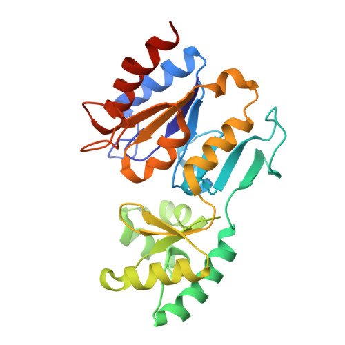
The Three-Dimensional Structure of Mannosyl-3-Phosphoglycerate Phosphatase from Thermus Thermophilus Hb27: A New Member of the Haloalkanoic Acid Dehalogenase Superfamily.

Goncalves, S.Esteves, A.M.Santos, H.Borges, N.Matias, P.M.

(2011) Biochemistry 50: 9551

  • DOI: https://doi.org/10.1021/bi201171h
  • Primary Citation of Related Structures:  
    3ZTW, 3ZTY, 3ZU6, 3ZUP, 3ZW7, 3ZWD, 3ZWK, 3ZX4, 3ZX5

  • PubMed Abstract: 

    Mannosyl-3-phosphoglycerate phosphatase (MpgP) is a key mediator in the physiological response to thermal and osmotic stresses, catalyzing the hydrolysis of mannosyl-3-phosphoglycerate (MPG) to the final product, α-mannosylglycerate. MpgP is a metal-dependent haloalcanoic acid dehalogenase-like (HAD-like) phosphatase, preserving the catalytic motifs I-IV of the HAD core domain, and classified as a Cof-type MPGP (HAD-IIB-MPGP family; SCOP [117505]) on the basis of its C2B cap insertion module. Herein, the crystallographic structures of Thermus thermophilus HB27 MpgP in its apo form and in complex with substrates, substrate analogues, and inhibitors are reported. Two distinct enzyme conformations, open and closed, are catalytically relevant. Apo-MpgP is primarily found in the open state, while holo-MpgP, in complex with the reaction products, is found in the closed state. Enzyme activation entails a structural rearrangement of motifs I and IV with concomitant binding of the cocatalytic Mg(2+) ion. The closure motion of the C2B domain is subsequently triggered by the anchoring of the phosphoryl group to the cocatalytic metal center, and by Arg167 fixing the mannosyl moiety inside the catalytic pocket. The results led to the proposal that in T. thermophilus HB27 MpgP the phosphoryl transfer employs a concerted D(N)S(N) mechanism with assistance of proton transfer from the general acid Asp8, forming a short-lived PO(3)(-) intermediate that is attacked by a nucleophilic water molecule. These results provide new insights into a possible continuum of phosphoryl transfer mechanisms, ranging between those purely associative and dissociative, as well as a picture of the main mechanistic aspects of phosphoryl monoester transfer catalysis, common to other members of the HAD superfamily.


  • Organizational Affiliation

    ITQB-Instituto de Tecnologia Quı́mica e Biológica, Universidade Nova de Lisboa, Apartado 127, 2781-901 Oeiras, Portugal.


Macromolecules
Find similar proteins by:  (by identity cutoff)  |  3D Structure
Entity ID: 1
MoleculeChains Sequence LengthOrganismDetailsImage
MANNOSYL-3-PHOSPHOGLYCERATE PHOSPHATASE
A, B
259Thermus thermophilus HB27Mutation(s): 0 
EC: 3.1.3.70
UniProt
Find proteins for Q72K29 (Thermus thermophilus (strain ATCC BAA-163 / DSM 7039 / HB27))
Explore Q72K29 
Go to UniProtKB:  Q72K29
Entity Groups  
Sequence Clusters30% Identity50% Identity70% Identity90% Identity95% Identity100% Identity
UniProt GroupQ72K29
Sequence Annotations
Expand
  • Reference Sequence
Small Molecules
Ligands 5 Unique
IDChains Name / Formula / InChI Key2D Diagram3D Interactions
2M8
Query on 2M8

Download Ideal Coordinates CCD File 
H [auth B](2R)-3-hydroxy-2-(alpha-D-mannopyranosyloxy)propanoic acid
C9 H16 O9
DDXCFDOPXBPUJC-SAYMMRJXSA-N
PO4
Query on PO4

Download Ideal Coordinates CCD File 
D [auth A]PHOSPHATE ION
O4 P
NBIIXXVUZAFLBC-UHFFFAOYSA-K
PO3
Query on PO3

Download Ideal Coordinates CCD File 
G [auth B]PHOSPHITE ION
O3 P
AQSJGOWTSHOLKH-UHFFFAOYSA-N
CL
Query on CL

Download Ideal Coordinates CCD File 
E [auth A]CHLORIDE ION
Cl
VEXZGXHMUGYJMC-UHFFFAOYSA-M
MG
Query on MG

Download Ideal Coordinates CCD File 
C [auth A],
F [auth B]
MAGNESIUM ION
Mg
JLVVSXFLKOJNIY-UHFFFAOYSA-N
Experimental Data & Validation

Experimental Data

  • Method: X-RAY DIFFRACTION
  • Resolution: 1.90 Å
  • R-Value Free: 0.203 
  • R-Value Work: 0.162 
  • R-Value Observed: 0.164 
  • Space Group: P 1 21 1
Unit Cell:
Length ( Å )Angle ( ˚ )
a = 39.794α = 90
b = 70.804β = 95.54
c = 92.77γ = 90
Software Package:
Software NamePurpose
PHENIXrefinement
XDSdata reduction
XDSdata scaling
PHASERphasing

Structure Validation

View Full Validation Report


Ligand Structure Quality Assessment 


Entry History 

Deposition Data

Revision History  (Full details and data files)

  • Version 1.0: 2011-10-19
    Type: Initial release
  • Version 1.1: 2011-11-09
    Changes: Database references
  • Version 2.0: 2020-07-29
    Type: Remediation
    Reason: Carbohydrate remediation
    Changes: Atomic model, Derived calculations, Other, Structure summary
  • Version 2.1: 2023-12-20
    Changes: Data collection, Database references, Refinement description, Structure summary