


Crystal Structure of the Human Glyoxalase Domain-Containing Protein 5
Muniz, J.R.C., Kiyani, W., Vollmar, M., von Delft, F., Burgess-Brown, N., Arrowsmith, C.H., Edwards, A.M., Weigelt, J., Bountra, C., Yue, W.W.To be published.
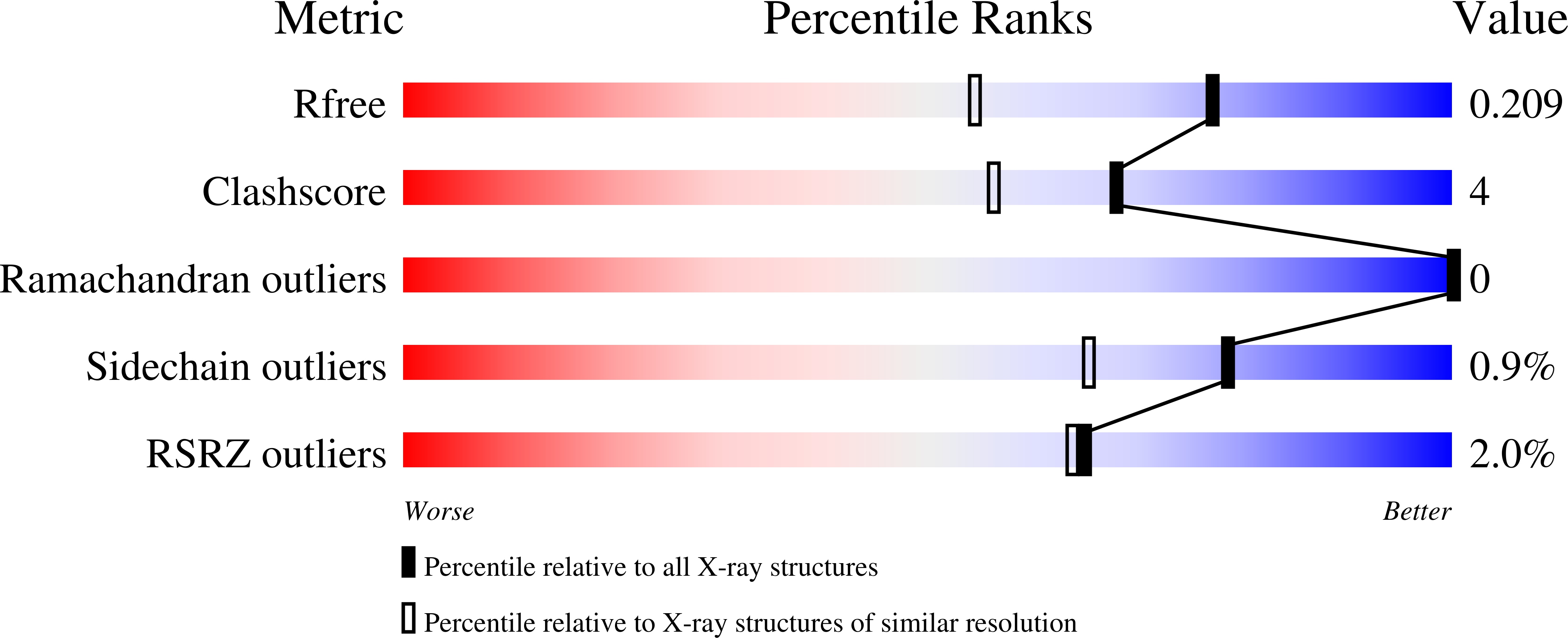
Experimental Data Snapshot
wwPDB Validation 3D Report Full Report
Entity ID: 1 | |||||
|---|---|---|---|---|---|
| Molecule | Chains | Sequence Length | Organism | Details | Image |
| GLYOXALASE DOMAIN-CONTAINING PROTEIN 5 | 147 | Homo sapiens | Mutation(s): 0 | 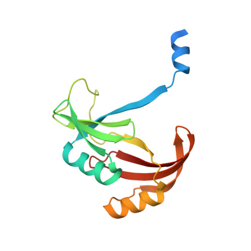 | |
UniProt & NIH Common Fund Data Resources | |||||
Find proteins for A6NK44 (Homo sapiens) Explore A6NK44 Go to UniProtKB: A6NK44 | |||||
PHAROS: A6NK44 GTEx: ENSG00000171433 | |||||
Entity Groups | |||||
| Sequence Clusters | 30% Identity50% Identity70% Identity90% Identity95% Identity100% Identity | ||||
| UniProt Group | A6NK44 | ||||
Sequence AnnotationsExpand | |||||
| |||||
| Ligands 3 Unique | |||||
|---|---|---|---|---|---|
| ID | Chains | Name / Formula / InChI Key | 2D Diagram | 3D Interactions | |
| PEG Query on PEG | D [auth A] | DI(HYDROXYETHYL)ETHER C4 H10 O3 MTHSVFCYNBDYFN-UHFFFAOYSA-N |  | ||
| SO4 Query on SO4 | C [auth A], F [auth B] | SULFATE ION O4 S QAOWNCQODCNURD-UHFFFAOYSA-L |  | ||
| EDO Query on EDO | E [auth A], G [auth B] | 1,2-ETHANEDIOL C2 H6 O2 LYCAIKOWRPUZTN-UHFFFAOYSA-N |  | ||
| Length ( Å ) | Angle ( ˚ ) |
|---|---|
| a = 51.49 | α = 90 |
| b = 66.04 | β = 90 |
| c = 76.87 | γ = 90 |
| Software Name | Purpose |
|---|---|
| BUSTER | refinement |
| MOSFLM | data reduction |
| SCALA | data scaling |
| PHASER | phasing |
RCSB PDB (citation) is hosted by
RCSB PDB is a member of the

