3ZVN

The structural basis for substrate recognition by mammalian polynucleotide kinase 3' phosphatase


Experimental Data Snapshot

  • Method: X-RAY DIFFRACTION
  • Resolution: 2.15 Å
  • R-Value Free: 0.269 
  • R-Value Work: 0.205 
  • R-Value Observed: 0.208 

wwPDB Validation   3D Report Full Report


Ligand Structure Quality Assessment 


This is version 1.2 of the entry. See complete history


Literature

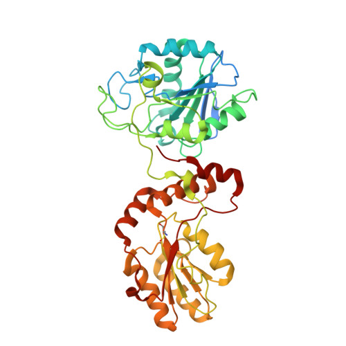
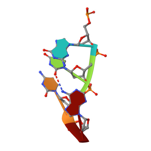
The structural basis for substrate recognition by mammalian polynucleotide kinase 3' phosphatase.

Garces, F.Pearl, L.H.Oliver, A.W.

(2011) Mol Cell 44: 385-396

  • DOI: https://doi.org/10.1016/j.molcel.2011.08.036
  • Primary Citation of Related Structures:  
    3ZVL, 3ZVM, 3ZVN

  • PubMed Abstract: 

    Mammalian polynucleotide kinase 3' phosphatase (PNK) plays a key role in the repair of DNA damage, functioning as part of both the nonhomologous end-joining (NHEJ) and base excision repair (BER) pathways. Through its two catalytic activities, PNK ensures that DNA termini are compatible with extension and ligation by either removing 3'-phosphates from, or by phosphorylating 5'-hydroxyl groups on, the ribose sugar of the DNA backbone. We have now determined crystal structures of murine PNK with DNA molecules bound to both of its active sites. The structure of ssDNA engaged with the 3'-phosphatase domain suggests a mechanism of substrate interaction that assists DNA end seeking. The structure of dsDNA bound to the 5'-kinase domain reveals a mechanism of DNA bending that facilitates recognition of DNA ends in the context of single-strand and double-strand breaks and suggests a close functional cooperation in substrate recognition between the kinase and phosphatase active sites.


  • Organizational Affiliation

    Cancer Research UK DNA Repair Enzymes Group, Genome Damage and Stability Centre, School of Life Sciences, University of Sussex, Falmer BN1 9QG, UK.


Macromolecules

Find similar proteins by:  (by identity cutoff)  |  3D Structure
Entity ID: 1
MoleculeChains Sequence LengthOrganismDetailsImage
BIFUNCTIONAL POLYNUCLEOTIDE PHOSPHATASE/KINASE416Mus musculusMutation(s): 1 
EC: 3.1.3.32 (PDB Primary Data), 2.7.1.78 (PDB Primary Data)
UniProt & NIH Common Fund Data Resources
Find proteins for Q9JLV6 (Mus musculus)
Explore Q9JLV6 
Go to UniProtKB:  Q9JLV6
IMPC:  MGI:1891698
Entity Groups  
Sequence Clusters30% Identity50% Identity70% Identity90% Identity95% Identity100% Identity
UniProt GroupQ9JLV6
Sequence Annotations
Expand
  • Reference Sequence

Find similar nucleic acids by:  Sequence   |   3D Structure  

Entity ID: 2
MoleculeChains LengthOrganismImage
5'-D(*GP*TP*CP*AP*CP)-3'B [auth E],
C [auth F],
D [auth G],
E [auth H],
F [auth I]
5synthetic construct
Sequence Annotations
Expand
  • Reference Sequence
Small Molecules
Experimental Data & Validation

Experimental Data

  • Method: X-RAY DIFFRACTION
  • Resolution: 2.15 Å
  • R-Value Free: 0.269 
  • R-Value Work: 0.205 
  • R-Value Observed: 0.208 
  • Space Group: P 21 21 21
Unit Cell:
Length ( Å )Angle ( ˚ )
a = 69.27α = 90
b = 72.99β = 90
c = 114.44γ = 90
Software Package:
Software NamePurpose
PHENIXrefinement
iMOSFLMdata reduction
SCALAdata scaling
PHASERphasing

Structure Validation

View Full Validation Report


Ligand Structure Quality Assessment 


Entry History 

Deposition Data

Revision History  (Full details and data files)

  • Version 1.0: 2011-11-16
    Type: Initial release
  • Version 1.1: 2018-03-07
    Changes: Database references, Source and taxonomy
  • Version 1.2: 2023-12-20
    Changes: Data collection, Database references, Derived calculations, Other, Refinement description