3ZOI

ISOPENICILLIN N SYNTHASE WITH AC-O-METHYL-D-THREONINE


Experimental Data Snapshot

  • Method: X-RAY DIFFRACTION
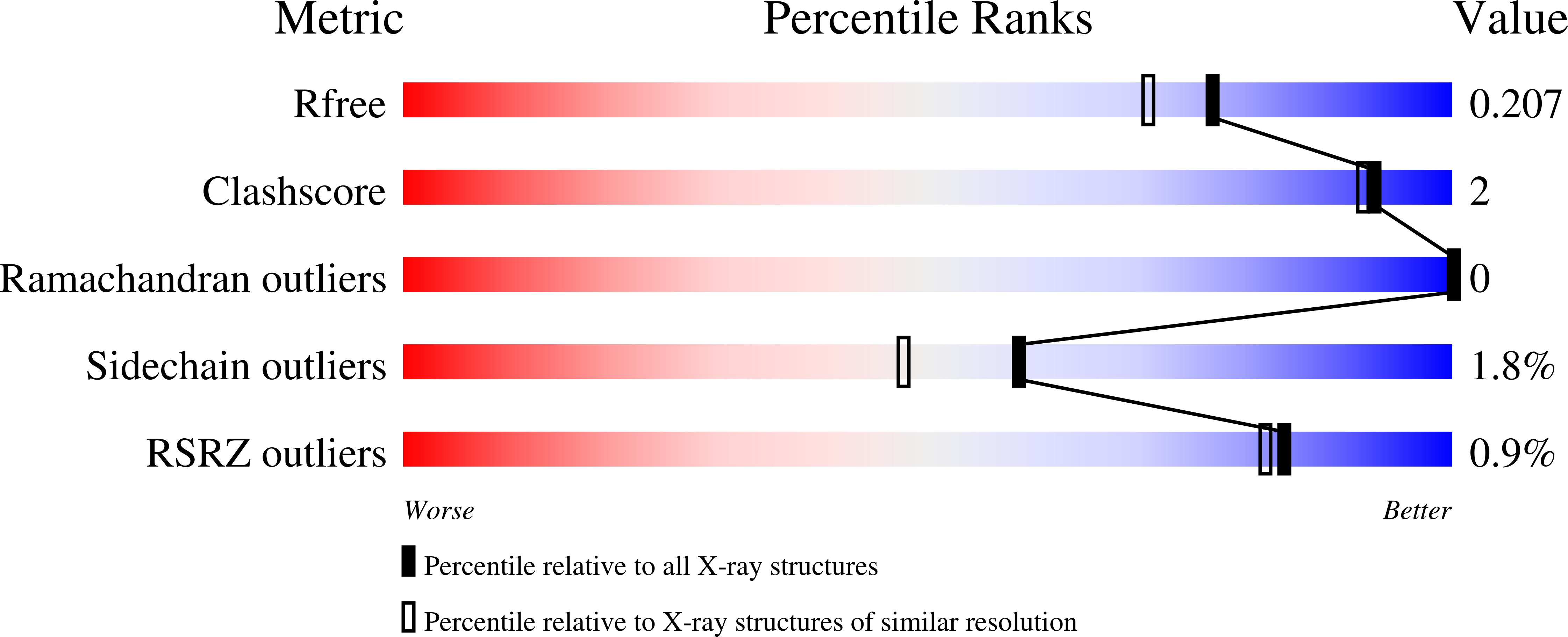
  • Resolution: 1.82 Å
  • R-Value Free: 0.202 
  • R-Value Work: 0.160 
  • R-Value Observed: 0.162 

wwPDB Validation   3D Report Full Report


Ligand Structure Quality Assessment 


This is version 1.3 of the entry. See complete history


Literature

The Crystal Structure of an Isopenicillin N Synthase Complex with an Ethereal Substrate Analogue Reveals Water in the Oxygen Binding Site.

Clifton, I.J.Ge, W.Adlington, R.M.Baldwin, J.E.Rutledge, P.J.

(2013) FEBS Lett 587: 2705

  • DOI: https://doi.org/10.1016/j.febslet.2013.07.016
  • Primary Citation of Related Structures:  
    3ZOI

  • PubMed Abstract: 

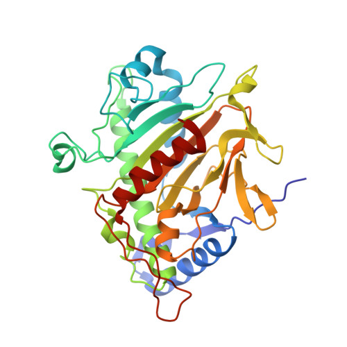
    Isopenicillin N synthase (IPNS) is a non-heme iron oxidase central to the biosynthesis of β-lactam antibiotics. IPNS converts the tripeptide δ-(L-α-aminoadipoyl)-L-cysteinyl-D-valine (ACV) to isopenicillin N while reducing molecular oxygen to water. The substrate analogue δ-(L-α-aminoadipoyl)-L-cysteinyl-O-methyl-D-threonine (ACmT) is not turned over by IPNS. Epimeric δ-(L-α-aminoadipoyl)-L-cysteinyl-O-methyl-D-allo-threonine (ACmaT) is converted to a bioactive penam product. ACmT and ACmaT differ from each other only in the stereochemistry at the β-carbon atom of their third residue. These substrates both contain a methyl ether in place of the isopropyl group of ACV. We report an X-ray crystal structure for the anaerobic IPNS:Fe(II):ACmT complex. This structure reveals an additional water molecule bound to the active site metal, held by hydrogen-bonding to the ether oxygen atom of the substrate analogue.


  • Organizational Affiliation

    Chemistry Research Laboratory, University of Oxford, Mansfield Road, Oxford OX1 3TA, UK.


Macromolecules
Find similar proteins by:  (by identity cutoff)  |  3D Structure
Entity ID: 1
MoleculeChains Sequence LengthOrganismDetailsImage
ISOPENICILLIN N SYNTHASE331Aspergillus nidulansMutation(s): 0 
EC: 1.21.3.1
UniProt
Find proteins for P05326 (Emericella nidulans (strain FGSC A4 / ATCC 38163 / CBS 112.46 / NRRL 194 / M139))
Explore P05326 
Go to UniProtKB:  P05326
Entity Groups  
Sequence Clusters30% Identity50% Identity70% Identity90% Identity95% Identity100% Identity
UniProt GroupP05326
Sequence Annotations
Expand
  • Reference Sequence
Small Molecules
Ligands 4 Unique
IDChains Name / Formula / InChI Key2D Diagram3D Interactions
M2W
Query on M2W

Download Ideal Coordinates CCD File 
B [auth A]DELTA-(L-ALPHA-AMINOADIPOYL)-L-CYSTEINYL-O-METHYL-D-THREONINE
C14 H25 N3 O7 S
LVCFAJQOVSJZGP-FTYOSLGDSA-N
SO4
Query on SO4

Download Ideal Coordinates CCD File 
D [auth A],
E [auth A],
F [auth A],
H [auth A]
SULFATE ION
O4 S
QAOWNCQODCNURD-UHFFFAOYSA-L
GOL
Query on GOL

Download Ideal Coordinates CCD File 
G [auth A]GLYCEROL
C3 H8 O3
PEDCQBHIVMGVHV-UHFFFAOYSA-N
FE
Query on FE

Download Ideal Coordinates CCD File 
C [auth A]FE (III) ION
Fe
VTLYFUHAOXGGBS-UHFFFAOYSA-N
Experimental Data & Validation

Experimental Data

  • Method: X-RAY DIFFRACTION
  • Resolution: 1.82 Å
  • R-Value Free: 0.202 
  • R-Value Work: 0.160 
  • R-Value Observed: 0.162 
  • Space Group: P 21 21 21
Unit Cell:
Length ( Å )Angle ( ˚ )
a = 46.48α = 90
b = 70.79β = 90
c = 100.51γ = 90
Software Package:
Software NamePurpose
REFMACrefinement
DENZOdata reduction
SCALAdata scaling

Structure Validation

View Full Validation Report


Ligand Structure Quality Assessment 


Entry History 

Deposition Data

Revision History  (Full details and data files)

  • Version 1.0: 2013-06-26
    Type: Initial release
  • Version 1.1: 2014-10-22
    Changes: Database references
  • Version 1.2: 2017-07-05
    Changes: Refinement description
  • Version 1.3: 2023-12-20
    Changes: Data collection, Database references, Derived calculations, Other, Refinement description