3ZLJ

CRYSTAL STRUCTURE OF FULL-LENGTH E.COLI DNA MISMATCH REPAIR PROTEIN MUTS D835R MUTANT IN COMPLEX WITH GT MISMATCHED DNA


Experimental Data Snapshot

  • Method: X-RAY DIFFRACTION
  • Resolution: 3.10 Å
  • R-Value Free: 0.263 
  • R-Value Work: 0.227 
  • R-Value Observed: 0.229 

wwPDB Validation   3D Report Full Report


This is version 1.4 of the entry. See complete history


Literature

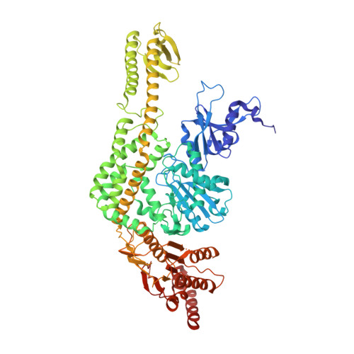
Using Stable Muts Dimers and Tetramers to Quantitatively Analyze DNA Mismatch Recognition and Sliding Clamp Formation.

Groothuizen, F.S.Fish, A.Petoukhov, M.V.Reumer, A.Manelyte, L.Winterwerp, H.H.K.Marinus, M.G.Lebbink, J.H.G.Svergun, D.I.Friedhoff, P.Sixma, T.K.

(2013) Nucleic Acids Res 41: 8166

  • DOI: https://doi.org/10.1093/nar/gkt582
  • Primary Citation of Related Structures:  
    3ZLJ

  • PubMed Abstract: 

    The process of DNA mismatch repair is initiated when MutS recognizes mismatched DNA bases and starts the repair cascade. The Escherichia coli MutS protein exists in an equilibrium between dimers and tetramers, which has compromised biophysical analysis. To uncouple these states, we have generated stable dimers and tetramers, respectively. These proteins allowed kinetic analysis of DNA recognition and structural analysis of the full-length protein by X-ray crystallography and small angle X-ray scattering. Our structural data reveal that the tetramerization domains are flexible with respect to the body of the protein, resulting in mostly extended structures. Tetrameric MutS has a slow dissociation from DNA, which can be due to occasional bending over and binding DNA in its two binding sites. In contrast, the dimer dissociation is faster, primarily dependent on a combination of the type of mismatch and the flanking sequence. In the presence of ATP, we could distinguish two kinetic groups: DNA sequences where MutS forms sliding clamps and those where sliding clamps are not formed efficiently. Interestingly, this inability to undergo a conformational change rather than mismatch affinity is correlated with mismatch repair.


  • Organizational Affiliation

    Division of Biochemistry and CancerGenomiCs.nl, Netherlands Cancer Institute, Plesmanlaan 121, 1066 CX Amsterdam, the Netherlands, European Molecular Biology Laboratory, Hamburg Outstation, EMBL c/o DESY, Notkestrasse 85, 22607 Hamburg, Germany, Institute for Biochemistry, Justus Liebig University, Heinrich-Buff Ring 58, D-35392, Giessen, Germany, Department of Biochemistry and Molecular Pharmacology, University of Massachusetts Medical School, 364 Plantation Street, Worcester, MA 01605, USA, Department of Cell Biology and Genetics, Cancer Genomics Center, Erasmus Medical Center, PO Box 2040, 3000 CA Rotterdam, the Netherlands and Department of Radiation Oncology, Erasmus Medical Center, PO Box 2040, 3000 CA Rotterdam, the Netherlands.


Macromolecules

Find similar proteins by:  (by identity cutoff)  |  3D Structure
Entity ID: 1
MoleculeChains Sequence LengthOrganismDetailsImage
DNA MISMATCH REPAIR PROTEIN MUTS
A, B
800Escherichia coli K-12Mutation(s): 0 
UniProt
Find proteins for P23909 (Escherichia coli (strain K12))
Explore P23909 
Go to UniProtKB:  P23909
Entity Groups  
Sequence Clusters30% Identity50% Identity70% Identity90% Identity95% Identity100% Identity
UniProt GroupP23909
Sequence Annotations
Expand
  • Reference Sequence
Find similar proteins by:  (by identity cutoff)  |  3D Structure
Entity ID: 2
MoleculeChains Sequence LengthOrganismDetailsImage
DNA MISMATCH REPAIR PROTEIN MUTS
C, D
53Escherichia coli K-12Mutation(s): 1 
UniProt
Find proteins for P23909 (Escherichia coli (strain K12))
Explore P23909 
Go to UniProtKB:  P23909
Entity Groups  
Sequence Clusters30% Identity50% Identity70% Identity90% Identity95% Identity100% Identity
UniProt GroupP23909
Sequence Annotations
Expand
  • Reference Sequence

Find similar nucleic acids by:  Sequence   |   3D Structure  

Entity ID: 3
MoleculeChains LengthOrganismImage
5'-D(*AP*GP*CP*TP*GP*CP*CP*AP*GP*GP*CP*AP*CP*CP *AP*GP*TP*GP*TP*CP*AP)-3'21synthetic construct
Sequence Annotations
Expand
  • Reference Sequence

Find similar nucleic acids by:  Sequence   |   3D Structure  

Entity ID: 4
MoleculeChains LengthOrganismImage
5'-D(*TP*GP*AP*CP*AP*CP*TP*GP*GP*TP*GP*CP*TP*TP *GP*GP*CP*AP*GP*CP*TP)-3'21synthetic construct
Sequence Annotations
Expand
  • Reference Sequence
Experimental Data & Validation

Experimental Data

  • Method: X-RAY DIFFRACTION
  • Resolution: 3.10 Å
  • R-Value Free: 0.263 
  • R-Value Work: 0.227 
  • R-Value Observed: 0.229 
  • Space Group: P 1 21 1
Unit Cell:
Length ( Å )Angle ( ˚ )
a = 110.288α = 90
b = 91.152β = 101.79
c = 112.855γ = 90
Software Package:
Software NamePurpose
REFMACrefinement
XDSdata reduction
SCALAdata scaling
PHASERphasing

Structure Validation

View Full Validation Report


Entry History 

Revision History  (Full details and data files)

  • Version 1.0: 2013-07-17
    Type: Initial release
  • Version 1.1: 2013-10-30
    Changes: Database references
  • Version 1.2: 2019-01-30
    Changes: Data collection, Experimental preparation, Other
  • Version 1.3: 2019-02-06
    Changes: Data collection, Experimental preparation
  • Version 1.4: 2023-12-20
    Changes: Data collection, Database references, Other, Refinement description