Stability of the Octameric Structure Affects Plasminogen-Binding Capacity of Streptococcal Enolase.
Cork, A.J., Ericsson, D.J., Law, R.H.P., Casey, L.W., Valkov, E., Bertozzi, C., Stamp, A., Jovcevski, B., Aquilina, J.A., Whisstock, J.C., Walker, M.J., Kobe, B.(2015) PLoS One 10: 21764
- PubMed: 25807546
- DOI: https://doi.org/10.1371/journal.pone.0121764
- Primary Citation of Related Structures:
3ZLF, 3ZLG, 3ZLH - PubMed Abstract:
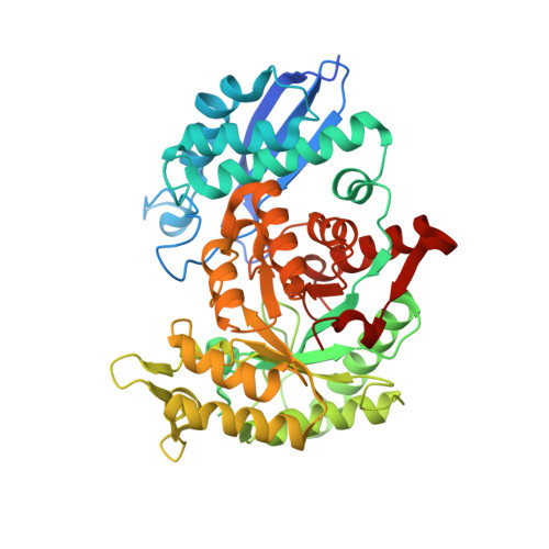
Group A Streptococcus (GAS) is a human pathogen that has the potential to cause invasive disease by binding and activating human plasmin(ogen). Streptococcal surface enolase (SEN) is an octameric α-enolase that is localized at the GAS cell surface. In addition to its glycolytic role inside the cell, SEN functions as a receptor for plasmin(ogen) on the bacterial surface, but the understanding of the molecular basis of plasmin(ogen) binding is limited. In this study, we determined the crystal and solution structures of GAS SEN and characterized the increased plasminogen binding by two SEN mutants. The plasminogen binding ability of SENK312A and SENK362A is ~2- and ~3.4-fold greater than for the wild-type protein. A combination of thermal stability assays, native mass spectrometry and X-ray crystallography approaches shows that increased plasminogen binding ability correlates with decreased stability of the octamer. We propose that decreased stability of the octameric structure facilitates the access of plasmin(ogen) to its binding sites, leading to more efficient plasmin(ogen) binding and activation.
Organizational Affiliation:
School of Chemistry and Molecular Biosciences and Institute for Molecular Bioscience, University of Queensland, Brisbane, QLD, 4072, Australia; Australian Infectious Disease Research Centre, University of Queensland, Brisbane, QLD, 4072, Australia.