3ZK1

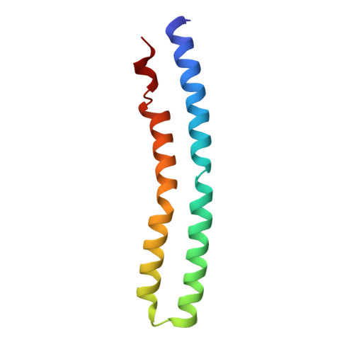
Crystal structure of the sodium binding rotor ring at pH 5.3


Experimental Data Snapshot

  • Method: X-RAY DIFFRACTION
  • Resolution: 2.20 Å
  • R-Value Free: 0.234 
  • R-Value Work: 0.208 
  • R-Value Observed: 0.209 

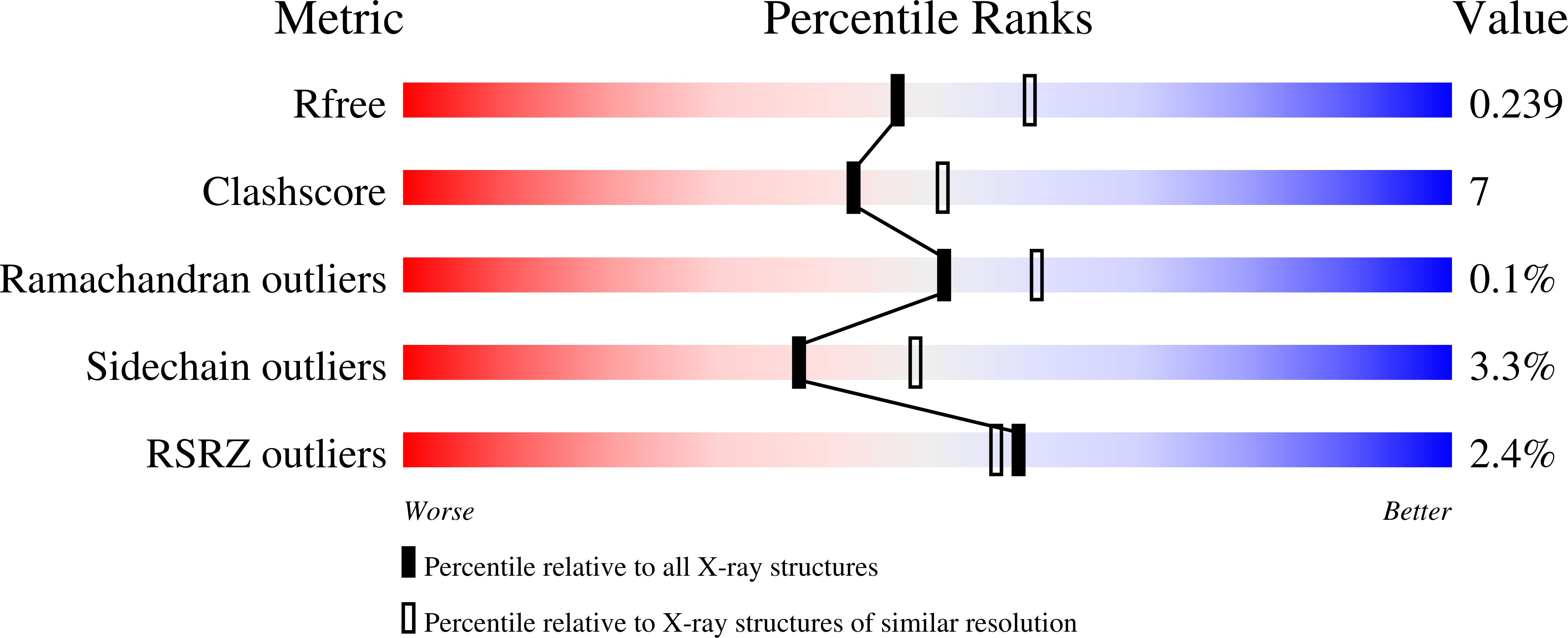
wwPDB Validation   3D Report Full Report


Ligand Structure Quality Assessment 


This is version 1.2 of the entry. See complete history


Literature

A New Type of Na(+)-Driven ATP Synthase Membrane Rotor with a Two-Carboxylate Ion-Coupling Motif.

Schulz, S.Iglesias-Cans, M.Krah, A.Yildiz, O.Leone, V.Matthies, D.Cook, G.M.Faraldo-Gomez, J.D.Meier, T.

(2013) PLoS Biol 11: 01596

  • DOI: https://doi.org/10.1371/journal.pbio.1001596
  • Primary Citation of Related Structures:  
    3ZK1, 3ZK2

  • PubMed Abstract: 

    The anaerobic bacterium Fusobacterium nucleatum uses glutamate decarboxylation to generate a transmembrane gradient of Na⁺. Here, we demonstrate that this ion-motive force is directly coupled to ATP synthesis, via an F₁F₀-ATP synthase with a novel Na⁺ recognition motif, shared by other human pathogens. Molecular modeling and free-energy simulations of the rotary element of the enzyme, the c-ring, indicate Na⁺ specificity in physiological settings. Consistently, activity measurements showed Na⁺ stimulation of the enzyme, either membrane-embedded or isolated, and ATP synthesis was sensitive to the Na⁺ ionophore monensin. Furthermore, Na⁺ has a protective effect against inhibitors targeting the ion-binding sites, both in the complete ATP synthase and the isolated c-ring. Definitive evidence of Na⁺ coupling is provided by two identical crystal structures of the c₁₁ ring, solved by X-ray crystallography at 2.2 and 2.6 Å resolution, at pH 5.3 and 8.7, respectively. Na⁺ ions occupy all binding sites, each coordinated by four amino acids and a water molecule. Intriguingly, two carboxylates instead of one mediate ion binding. Simulations and experiments demonstrate that this motif implies that a proton is concurrently bound to all sites, although Na⁺ alone drives the rotary mechanism. The structure thus reveals a new mode of ion coupling in ATP synthases and provides a basis for drug-design efforts against this opportunistic pathogen.


  • Organizational Affiliation

    Department of Structural Biology, Max Planck Institute of Biophysics, Frankfurt am Main, Germany.


Macromolecules
Find similar proteins by:  (by identity cutoff)  |  3D Structure
Entity ID: 1
MoleculeChains Sequence LengthOrganismDetailsImage
ATP SYNTHASE SUBUNIT C
A, B, C, D, E
A, B, C, D, E, F, G, H, I, J, K, L, M, N, O, P, Q, R, S, T, U, V
89Fusobacterium nucleatumMutation(s): 0 
Membrane Entity: Yes 
UniProt
Find proteins for Q8RGD7 (Fusobacterium nucleatum subsp. nucleatum (strain ATCC 25586 / DSM 15643 / BCRC 10681 / CIP 101130 / JCM 8532 / KCTC 2640 / LMG 13131 / VPI 4355))
Explore Q8RGD7 
Go to UniProtKB:  Q8RGD7
Entity Groups  
Sequence Clusters30% Identity50% Identity70% Identity90% Identity95% Identity100% Identity
UniProt GroupQ8RGD7
Sequence Annotations
Expand
  • Reference Sequence
Small Molecules
Ligands 4 Unique
IDChains Name / Formula / InChI Key2D Diagram3D Interactions
LMT
Query on LMT

Download Ideal Coordinates CCD File 
AA [auth B],
DA [auth C],
EA [auth C]
DODECYL-BETA-D-MALTOSIDE
C24 H46 O11
NLEBIOOXCVAHBD-QKMCSOCLSA-N
DMU
Query on DMU

Download Ideal Coordinates CCD File 
BB [auth N]
CA [auth C]
DB [auth O]
FB [auth P]
GA [auth D]
BB [auth N],
CA [auth C],
DB [auth O],
FB [auth P],
GA [auth D],
HB [auth Q],
IA [auth E],
JB [auth R],
KA [auth F],
MA [auth G],
MB [auth S],
OA [auth H],
OB [auth T],
QA [auth I],
QB [auth U],
SA [auth J],
SB [auth V],
UA [auth K],
WA [auth L],
X [auth A],
YA [auth M],
Z [auth B]
DECYL-BETA-D-MALTOPYRANOSIDE
C22 H42 O11
WOQQAWHSKSSAGF-WXFJLFHKSA-N
TAM
Query on TAM

Download Ideal Coordinates CCD File 
KB [auth R],
ZA [auth M]
TRIS(HYDROXYETHYL)AMINOMETHANE
C7 H17 N O3
GKODZWOPPOTFGA-UHFFFAOYSA-N
NA
Query on NA

Download Ideal Coordinates CCD File 
AB [auth N]
BA [auth C]
CB [auth O]
EB [auth P]
FA [auth D]
AB [auth N],
BA [auth C],
CB [auth O],
EB [auth P],
FA [auth D],
GB [auth Q],
HA [auth E],
IB [auth R],
JA [auth F],
LA [auth G],
LB [auth S],
NA [auth H],
NB [auth T],
PA [auth I],
PB [auth U],
RA [auth J],
RB [auth V],
TA [auth K],
VA [auth L],
W [auth A],
XA [auth M],
Y [auth B]
SODIUM ION
Na
FKNQFGJONOIPTF-UHFFFAOYSA-N
Experimental Data & Validation

Experimental Data

  • Method: X-RAY DIFFRACTION
  • Resolution: 2.20 Å
  • R-Value Free: 0.234 
  • R-Value Work: 0.208 
  • R-Value Observed: 0.209 
  • Space Group: P 1 21 1
Unit Cell:
Length ( Å )Angle ( ˚ )
a = 135.77α = 90
b = 83.9β = 112.85
c = 150.99γ = 90
Software Package:
Software NamePurpose
PHENIXrefinement
XDSdata reduction
XSCALEdata scaling
PHASERphasing

Structure Validation

View Full Validation Report


Ligand Structure Quality Assessment 


Entry History 

Deposition Data

Revision History  (Full details and data files)

  • Version 1.0: 2013-05-29
    Type: Initial release
  • Version 1.1: 2013-09-18
    Changes: Database references, Other
  • Version 1.2: 2023-12-20
    Changes: Data collection, Database references, Derived calculations, Other, Refinement description