3X1D

Crystal Structure of Atlastin from Drosophila melanogaster


Experimental Data Snapshot

  • Method: X-RAY DIFFRACTION
  • Resolution: 2.87 Å
  • R-Value Free: 0.379 
  • R-Value Work: 0.273 
  • R-Value Observed: 0.278 

wwPDB Validation   3D Report Full Report


Ligand Structure Quality Assessment 


This is version 1.4 of the entry. See complete history


Literature

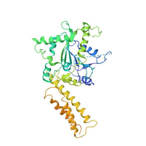
Comparison of human and Drosophila atlastin GTPases

Wu, F.Hu, X.Bian, X.Liu, X.Hu, J.

(2015) Protein Cell 6: 139-146

  • DOI: https://doi.org/10.1007/s13238-014-0118-0
  • Primary Citation of Related Structures:  
    3X1D

  • PubMed Abstract: 

    Formation of the endoplasmic reticulum (ER) network requires homotypic membrane fusion, which involves a class of atlastin (ATL) GTPases. Purified Drosophila ATL is capable of mediating vesicle fusion in vitro, but such activity has not been reported for any other ATLs. Here, we determined the preliminary crystal structure of the cytosolic segment of Drosophila ATL in a GDP-bound state. The structure reveals a GTPase domain dimer with the subsequent three-helix bundles associating with their own GTPase domains and pointing in opposite directions. This conformation is similar to that of human ATL1, to which GDP and high concentrations of inorganic phosphate, but not GDP only, were included. Drosophila ATL restored ER morphology defects in mammalian cells lacking ATLs, and measurements of nucleotide-dependent dimerization and GTPase activity were comparable for Drosophila ATL and human ATL1. However, purified and reconstituted human ATL1 exhibited no in vitro fusion activity. When the cytosolic segment of human ATL1 was connected to the transmembrane (TM) region and C-terminal tail (CT) of Drosophila ATL, the chimera still exhibited no fusion activity, though its GTPase activity was normal. These results suggest that GDP-bound ATLs may adopt multiple conformations and the in vitro fusion activity of ATL cannot be achieved by a simple collection of functional domains.


  • Organizational Affiliation

    Department of Genetics and Cell Biology, College of Life Sciences, Nankai University, Tianjin, 300071, China.


Macromolecules
Find similar proteins by:  (by identity cutoff)  |  3D Structure
Entity ID: 1
MoleculeChains Sequence LengthOrganismDetailsImage
Atlastin541Drosophila melanogasterMutation(s): 0 
Gene Names: atlCG6668
EC: 3.6.5
UniProt
Find proteins for Q9VC57 (Drosophila melanogaster)
Explore Q9VC57 
Go to UniProtKB:  Q9VC57
Entity Groups  
Sequence Clusters30% Identity50% Identity70% Identity90% Identity95% Identity100% Identity
UniProt GroupQ9VC57
Sequence Annotations
Expand
  • Reference Sequence
Small Molecules
Ligands 2 Unique
IDChains Name / Formula / InChI Key2D Diagram3D Interactions
GDP
Query on GDP

Download Ideal Coordinates CCD File 
B [auth A]GUANOSINE-5'-DIPHOSPHATE
C10 H15 N5 O11 P2
QGWNDRXFNXRZMB-UUOKFMHZSA-N
MG
Query on MG

Download Ideal Coordinates CCD File 
C [auth A]MAGNESIUM ION
Mg
JLVVSXFLKOJNIY-UHFFFAOYSA-N
Experimental Data & Validation

Experimental Data

  • Method: X-RAY DIFFRACTION
  • Resolution: 2.87 Å
  • R-Value Free: 0.379 
  • R-Value Work: 0.273 
  • R-Value Observed: 0.278 
  • Space Group: C 1 2 1
Unit Cell:
Length ( Å )Angle ( ˚ )
a = 71.617α = 90
b = 63.907β = 101.6
c = 107.762γ = 90
Software Package:
Software NamePurpose
HKL-2000data collection
PHASESphasing
PHENIXrefinement
HKL-2000data reduction
HKL-2000data scaling

Structure Validation

View Full Validation Report


Ligand Structure Quality Assessment 


Entry History 

Deposition Data

  • Released Date: 2015-10-28 
  • Deposition Author(s): Liu, X.

Revision History  (Full details and data files)

  • Version 1.0: 2015-10-28
    Type: Initial release
  • Version 1.1: 2018-01-31
    Changes: Experimental preparation
  • Version 1.2: 2018-08-08
    Changes: Data collection
  • Version 1.3: 2024-03-20
    Changes: Data collection, Database references, Derived calculations
  • Version 1.4: 2024-04-03
    Changes: Refinement description