


Allosteric D-lactate dehydrogenases from three Gram-negative bacteria
Furukawa, N., Togawa, M., Miyanaga, A., Nakajima, M., Taguchi, H.To be published.
Experimental Data Snapshot
wwPDB Validation 3D Report Full Report

Entity ID: 1 | |||||
|---|---|---|---|---|---|
| Molecule | Chains | Sequence Length | Organism | Details | Image |
| D-isomer specific 2-hydroxyacid dehydrogenase NAD-binding | 345 | Escherichia coli BL21(DE3) | Mutation(s): 0 Gene Names: B21_01364, ECBD_2243, ECD_01352, ldhA EC: 1.1.1.28 | 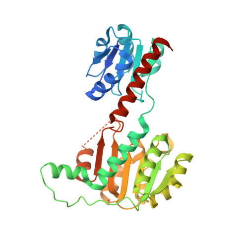 | |
Entity Groups | |||||
| Sequence Clusters | 30% Identity50% Identity70% Identity90% Identity95% Identity100% Identity | ||||
Sequence AnnotationsExpand | |||||
| |||||
| Length ( Å ) | Angle ( ˚ ) |
|---|---|
| a = 131.029 | α = 90 |
| b = 131.029 | β = 90 |
| c = 405.746 | γ = 120 |
| Software Name | Purpose |
|---|---|
| ADSC | data collection |
| MOLREP | phasing |
| REFMAC | refinement |
| MOSFLM | data reduction |
| SCALA | data scaling |
RCSB PDB (citation) is hosted by
RCSB PDB is a member of the

